. 2015:2015:301292.
doi: 10.1155/2015/301292. Epub 2015 Jan 5.

Prokineticin 2 upregulation in the peripheral nervous system has a major role in triggering and maintaining neuropathic pain in the chronic constriction injury model

Affiliations

Prokineticin 2 upregulation in the peripheral nervous system has a major role in triggering and maintaining neuropathic pain in the chronic constriction injury model

Roberta Lattanzi et al. Biomed Res Int. 2015.

Abstract

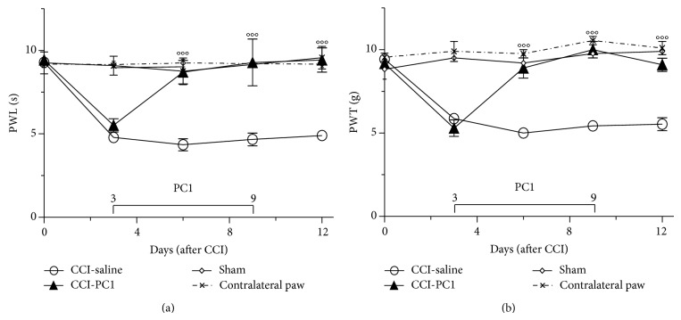
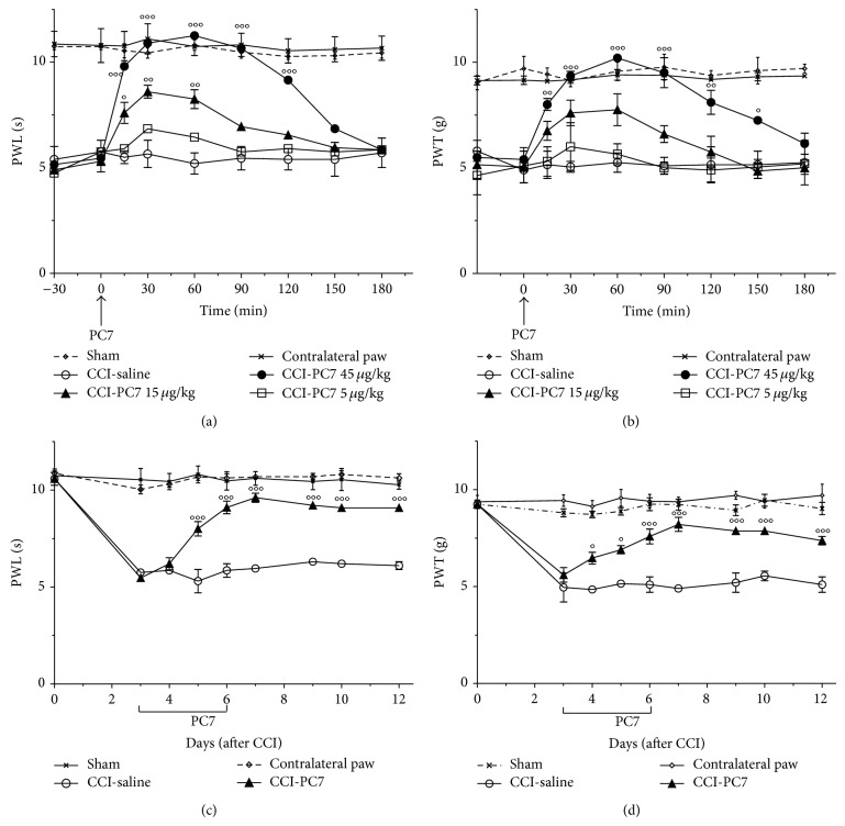
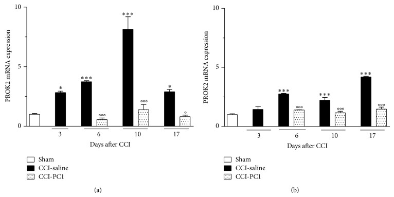
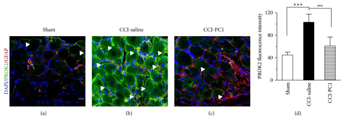
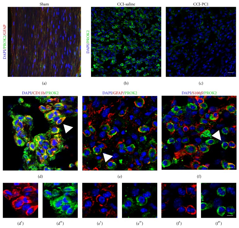
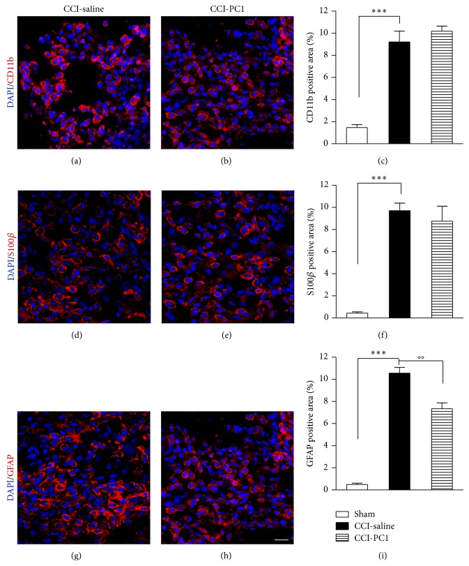
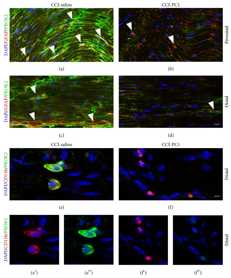
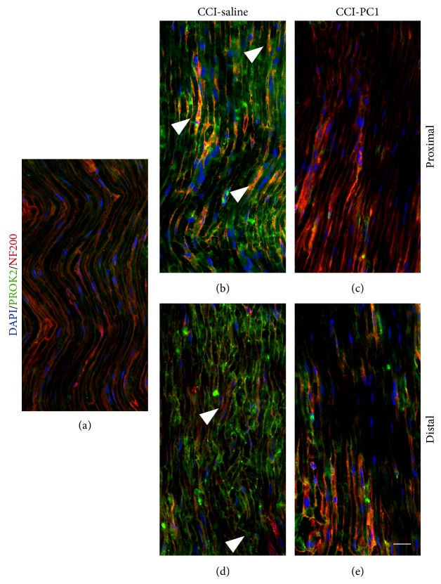
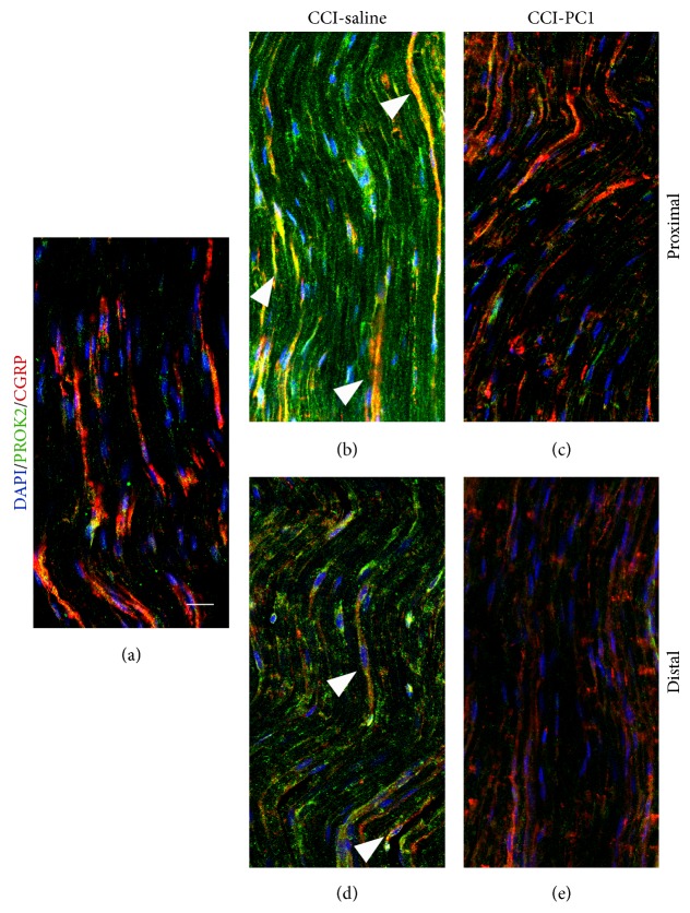
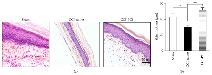
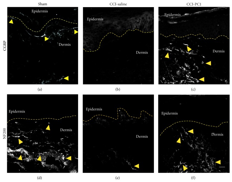
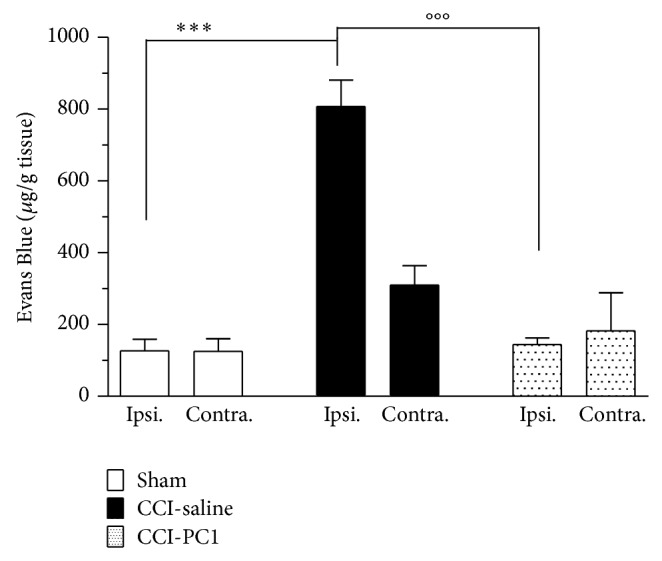
The new chemokine Prokineticin 2 (PROK2) and its receptors (PKR1 and PKR2) have a role in inflammatory pain and immunomodulation. Here we identified PROK2 as a critical mediator of neuropathic pain in the chronic constriction injury (CCI) of the sciatic nerve in mice and demonstrated that blocking the prokineticin receptors with two PKR1-preferring antagonists (PC1 and PC7) reduces pain and nerve damage. PROK2 mRNA expression was upregulated in the injured nerve since day 3 post injury (dpi) and in the ipsilateral DRG since 6 dpi. PROK2 protein overexpression was evident in Schwann Cells, infiltrating macrophages and axons in the peripheral nerve and in the neuronal bodies and some satellite cells in the DRG. Therapeutic treatment of neuropathic mice with the PKR-antagonist, PC1, impaired the PROK2 upregulation and signalling. This fact, besides alleviating pain, brought down the burden of proinflammatory cytokines in the damaged nerve and prompted an anti-inflammatory repair program. Such a treatment also reduced intraneural oedema and axon degeneration as demonstrated by the physiological skin innervation and thickness conserved in CCI-PC1 mice. These findings suggest that PROK2 plays a crucial role in neuropathic pain and might represent a novel target of treatment for this disease.


Figures

Figure 1
Figure 1
Repeated systemic injections of PC1 (150 μg kg−1, twice a day) from 3 to 9 dpi reverted the CCI-induced thermal hyperalgesia (a) and mechanical allodynia (b) in two days. The antihyperalgesic effect lasted after treatment withdrawal, for all the observation period. Data represent means ± SEM of 6–9 mice. Two-way ANOVA was used for statistical evaluation, followed by Bonferroni's test. °°°P < 0.001 CCI-PC1 versus CCI-saline mice.
Figure 2
Figure 2
Antihyperalgesic effect of PC7. A single bolus s.c. injection of PC7 (5, 15, and 45 μg kg−1) on day 3 after CCI dose-dependently reverted the established CCI-induced thermal hyperalgesia (a) and mechanical allodynia (b). The highest dose of PC7 (45 μg kg−1) abolished hyperalgesia for about 3 h. Repeated systemic injections of PC7 (45 μg kg−1, twice a day) from 3 to 7 dpi significantly reduced the CCI-induced thermal hyperalgesia (c) and mechanical allodynia (d) for all the observation period. Data represent means ± SEM of 5 mice. Two-way ANOVA was used for statistical evaluation, followed by Bonferroni's test. °P < 0.05; °°P < 0.01; °°°P < 0.001 CCI-PC7 versus CCI-saline mice.
Figure 3
Figure 3
Time-course of PROK2 mRNA expression in injured sciatic nerve and ipsilateral DRG of CCI-saline mice and CCI-PC1 mice. PROK2 levels in the peripheral nervous system of healthy animals were negligible (33.49 ± 0.09 Ct in sciatic nerve, 33.97 ± 0.41 Ct in DRG). In the sciatic nerve (a) PROK2 expression was significantly increased at 3 dpi, reached its maximal expression at 10 dpi, and then started to decrease up to 17 dpi. In the ipsilateral L 4–6 DRGs (b) PROK2 mRNA was significantly increased at 6 dpi and showed a constant tendency to increase up to 17 dpi. Data are mean ± SEM of 5 animals. One-way ANOVA was used for statistical evaluation, followed by Tukey test for multiple comparisons. * P < 0.05, *** P < 0.001 CCI-saline versus sham; °P < 0.05 CCI-PC1 versus CCI-saline.
Figure 4
Figure 4
Representative sections of mouse L4-L5 ipsilateral DRG, at 10 dpi, from sham (a), CCI-saline (b), and CCI-PC1 (c) mice. Immunofluorescence double-staining of PROK2 (green) with GFAP (marker for satellite cells, red). Cell nuclei were counterstained with DAPI (blue fluorescence). In DRG of sham-operated mice the PROK2 signal was very faint, localized only along cell membrane of some neurons, mainly small sized (arrowheads), and in few GFAP+ satellite cells (arrow) (a). In neurons of CCI-saline mice, PROK2 immunofluorescence was significantly increased and showed a vesicular cytoplasmatic pattern which is dense in proximity of the neuronal membrane (arrowheads). The number of PROK2+ satellite cells is increased (arrows). PROK2 signal in CCI-PC1 mice was comparable with that of sham mice (c). Scale bar, 30 μm. (d) Evaluation of PROK2 fluorescence intensity. One-way ANOVA was used for statistical evaluation, followed by Tukey test for multiple comparisons *** P < 0.001 CCI-saline versus sham mice; °°°P < 0.001 CCI-PC1 versus CCI-saline mice.
Figure 5
Figure 5
Representative images of sciatic nerve section in the immediate proximity of the injury, from sham (a), CCI-saline (b), and CCI-PC1 (c) mice at 10 dpi. (a) In the sciatic nerve of sham-operated mice PROK2 immunoreactivity (green) was very faint and colocalized with GFAP (red) in elongated SC. (b) A heavy infiltration of PROK2-positive cells (green) was evident in the nerve from CCI-saline mice. (c) PC1 treatment significantly reduced the PROK2 immunoreactivity in these cells. Scale bar, 30 μm. Immunofluorescence double-staining showing colocalization (yellow, arrowheads) of PROK2 (green) with CD11b (macrophage marker, red) (d), GFAP (Schwann cell marker, red) (e), and S100β (Schwann cell marker, red) (f) in the immediate proximity of the injury in the sciatic nerve of CCI-saline mice. (d′) CD11b (red), (e′) GFAP (red), (f′) S100β (red), and (d′′, e′′, f′′) PROK2 (green) shown in single channels. Scale bar, 10 μm. Cell nuclei were counterstained with DAPI (blue fluorescence).
Figure 6
Figure 6
Immunostaining of activated macrophages (CD11b+, red), S100β+ SC (red), and GFAP+ SC (red) in the neuroma from CCI-saline and CCI-PC1 mice ((a)–(h)). Repeated treatment with the PKR-antagonist significantly reduced the GFAP+ activated SC (i) but did not affect S100β+ SC or macrophage infiltration ((c), (f)).
Figure 7
Figure 7
Representative images of CCI-induced upregulation of PROK2 in the longitudinally sliced sciatic nerve proximal and distal to the lesion. At 10 dpi a dramatic increase of PROK2 signal (green, a and c) in fibres and in GFAP+ structures (yellow) was evident both proximal and distal to the lesion. The PROK2 signal was dramatically reduced in the nerve from CCI-PC1 mice (green, b and d). Scale bar: 30 μm. High-magnification images showed macrophages that infiltrate the nerve distal to the lesion (scale bar: 10 μm). (e) Double immunofluorescence labelling for PROK2 (green) and CD11b (red) showing that in the CCI-saline mice the infiltrating macrophages contain PROK2 (yellow). (f) PROK2 signal was absent in macrophages infiltrating the nerve from CCI-PC1 mice. (e′, f′) CD11b (red), and (e′′, f′′) PROK2 (green) shown in single channels. Cell nuclei were counterstained with DAPI (blue fluorescence).
Figure 8
Figure 8
Confocal images of representative sections of longitudinally sliced sciatic nerve, proximal and distal to the lesion, immunostained for PROK2 (green) and NF200 (red) from sham-operated, CCI-saline and CCI-PC1 mice at 10 dpi. Scale bar: 30 μm. PROK2-green signal was localized between NF200 positive fibers in CCI-saline mice but was not found in the nerve from CCI-PC1 mice.
Figure 9
Figure 9
Confocal images of representative sections of longitudinally sliced sciatic nerve proximal and distal to the lesion, immunostained for PROK2 (green) and CGRP (red) from sham-operated, CCI-saline and CCI-PC1 mice at 10 dpi. Scale bar: 30 μm.
Figure 10
Figure 10
(a) Histological examination of the plantar skin from sham, CCI-saline, and CCI-PC1 mice stained with hematoxylin-eosin at 10 dpi. (b) Quantification of epidermal thickness of the sham, CCI/saline, and CCI/PC1 mice (3 section/animal). Data are expressed as mean ± SEM of 4-5 animals. One-way ANOVA was used for statistical evaluation, followed by Tukey test for multiple comparisons: * P < 0.05; °°°P < 0.001.
Figure 11
Figure 11
Confocal images of representative skin sections immunostained for CGRP and NF200 from sham-operated, CCI-saline, and CCI-PC1 mice at 10 dpi. (a) In sham-operated mice the CGRP positive fibers were distributed along the dermoepidermal junction. (b) In CCI-saline mice the CGRP positive fibers were absent. (c) In CCI-PC1 mice the CGRP positive fibers were present in dermis. (d) In sham-operated mice the NF200 positive fibers were distributed along the dermoepidermal junction and in dermis. (e) In CCI-saline mice very few NF200 positive fibers were observed. (f) In CCI-PC1 mice the NF200 positive fibers were present in dermis. Dashed line represents the dermoepidermal junction. Scale bar 20 μm.
Figure 12
Figure 12
Evans Blue extravasation was measured at 3 dpi in injured and contralateral sciatic nerves. A significant increase in Evans Blue accumulation was evident in the injured nerve. The level of Evans Blue in the contralateral nerve was not significantly different from the level found in sham animals. Evans blue extravasation in the sciatic nerves of mice treated with PC1 (150 μg Kg−1, twice/day, at 1 and 2 dpi) did not differ from sham mice. Data are expressed as mean ± SEM of 4-5 animals. One-way ANOVA was used for statistical evaluation, followed by Tukey test for multiple comparisons: *** P < 0.001 CCI-saline versus sham mice; °°°P < 0.001 CCI-PC1 versus CCI-saline mice.

Similar articles

Cited by

References

    1. Abbadie C. Chemokines, chemokine receptors and pain. Trends in Immunology. 2005;26(10):529–534. doi: 10.1016/j.it.2005.08.001. - DOI - PubMed
    1. Negri L., Lattanzi R. Bv8/PK2 and prokineticin receptors: a druggable pronociceptive system. Current Opinion in Pharmacology. 2012;12(1):62–66. doi: 10.1016/j.coph.2011.10.023. - DOI - PubMed
    1. Giannini E., Lattanzi R., Nicotra A., et al. The chemokine Bv8/prokineticin 2 is up-regulated in inflammatory granulocytes and modulates inflammatory pain. Proceedings of the National Academy of Sciences of the United States of America. 2009;106(34):14646–14651. doi: 10.1073/pnas.0903720106. - DOI - PMC - PubMed
    1. LeCouter J., Zlot C., Tejada M., Peale F., Ferrara N. Bv8 and endocrine gland-derived vascular endothelial growth factor stimulate hematopoiesis and hematopoietic cell mobilization. Proceedings of the National Academy of Sciences of the United States of America. 2004;101(48):16813–16818. doi: 10.1073/pnas.0407697101. - DOI - PMC - PubMed
    1. Dorsch M., Qiu Y., Soler D., et al. PK1/EG-VEGF induces monocyte differentiation and activation. Journal of Leukocyte Biology. 2005;78(2):426–434. doi: 10.1189/jlb.0205061. - DOI - PubMed

Publication types