

Breast MRI in patients with unilateral bloody and serous-bloody nipple discharge: a comparison with galactography
- PMID: 25685810
- PMCID: PMC4317598
- DOI: 10.1155/2015/806368
Breast MRI in patients with unilateral bloody and serous-bloody nipple discharge: a comparison with galactography
Abstract
Purpose: Assessing the role of breast MRI compared to galactography in patients with unilateral bloody or serous-bloody nipple discharge.
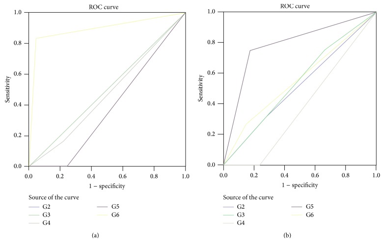
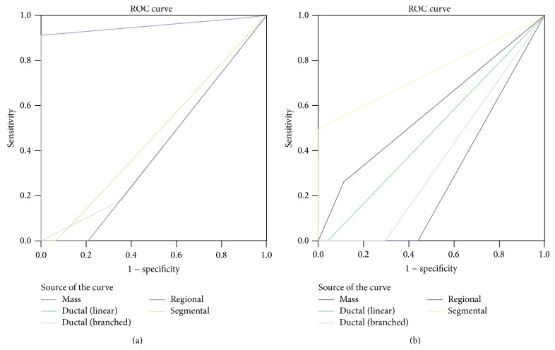
Materials and methods: Retrospective study including 53 unilateral discharge patients who performed galactography and MRI. We evaluated the capability of both techniques in identifying pathology and distinguishing between nonmalignant and malignant lesions. Lesions BIRADS 1/2 underwent follow-up, while the histological examination after surgery has been the gold standard to assess pathology in lesions BIRADS 3/4/5. The ROC analysis was used to test diagnostic MRI and galactography ability.
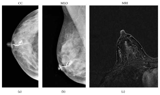
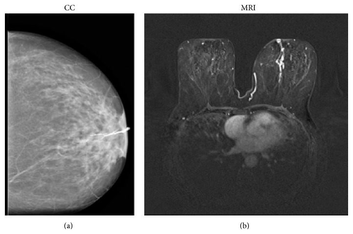
Results: After surgery and follow-up, 8 patients had no disease (15%), 23 papilloma (43%), 11 papillomatosis (21%), 5 ductal cancer in situ (10%), and 6 papillary carcinoma (11%) diagnoses. Both techniques presented 100% specificity; MRI sensitivity was 98% versus 49% of galactography. Considering MRI, we found a statistical association between mass enhancement and papilloma (P < 0.001; AUC 0.957; CI 0.888-1.025), ductal enhancement and papillomatosis (P < 0.001; AUC 0.790; CI 0.623-0.958), segmental enhancement and ductal cancer in situ (P = 0.007; AUC 0.750; CI 0.429-1.071), and linear enhancement and papillary cancer (P = 0.011).
Conclusions: MRI is a valid tool to detect ductal pathologies in patients with suspicious bloody or serous-bloody discharge showing higher sensitivity and specificity compared to galactography.
Figures
Similar articles
-
The diagnostic value of galactography in patients with nipple discharge.Clin Imaging. 2001 Mar-Apr;25(2):75-81. doi: 10.1016/s0899-7071(01)00256-x. Clin Imaging. 2001. PMID: 11483413
-
Preoperative galactography increases the diagnostic yield of major duct excision for nipple discharge.Cancer. 1998 May 15;82(10):1874-80. doi: 10.1002/(sici)1097-0142(19980515)82:10<1874::aid-cncr9>3.3.co;2-o. Cancer. 1998. PMID: 9587119
-
Role of breast magnetic resonance imaging (MRI) in patients with unilateral nipple discharge: preliminary study.Radiol Med. 2008 Mar;113(2):249-64. doi: 10.1007/s11547-008-0245-x. Epub 2008 Apr 2. Radiol Med. 2008. PMID: 18386126 English, Italian.
-
Management of bloody nipple discharge.Curr Treat Options Oncol. 2002 Apr;3(2):157-61. doi: 10.1007/s11864-002-0061-9. Curr Treat Options Oncol. 2002. PMID: 12057078 Review.
-
[Management of breast nipple discharge: Recommendations].J Gynecol Obstet Biol Reprod (Paris). 2015 Dec;44(10):927-37. doi: 10.1016/j.jgyn.2015.09.035. Epub 2015 Nov 3. J Gynecol Obstet Biol Reprod (Paris). 2015. PMID: 26545854 Review. French.
Cited by
-
Multiple Papillomas of the Breast: A Review of Current Evidence and Challenges.J Imaging. 2022 Jul 13;8(7):198. doi: 10.3390/jimaging8070198. J Imaging. 2022. PMID: 35877642 Free PMC article. Review.
-
Diagnostic Performance of Breast Magnetic Resonance Imaging in Non-Calcified Equivocal Breast Findings: Results from a Systematic Review and Meta-Analysis.PLoS One. 2016 Aug 2;11(8):e0160346. doi: 10.1371/journal.pone.0160346. eCollection 2016. PLoS One. 2016. PMID: 27482715 Free PMC article.
-
Understanding indications and defining guidelines for breast magnetic resonance imaging.SA J Radiol. 2018 Oct 30;22(2):1353. doi: 10.4102/sajr.v22i2.1353. eCollection 2018. SA J Radiol. 2018. PMID: 31754513 Free PMC article. Review.
-
The role of magnetic resonance imaging in detection and surgical treatment of breast intraductal papillomas.Transl Cancer Res. 2019 Apr;8(2):635-646. doi: 10.21037/tcr.2019.03.27. Transl Cancer Res. 2019. PMID: 35116796 Free PMC article.
-
Contribution of Diffusion-Weighted Imaging and ADC Values to Papillary Breast Lesions.Front Oncol. 2022 Jun 30;12:911790. doi: 10.3389/fonc.2022.911790. eCollection 2022. Front Oncol. 2022. PMID: 35847891 Free PMC article.
References
-
- Newman H. F., Klein M., Northrup J. D., Ray B. F., Drucker M. Nipple discharge: frequency and pathogenesis in an ambulatory population. New York State Journal of Medicine. 1983;83(7):928–933. - PubMed
Publication types
MeSH terms
LinkOut - more resources
Full Text Sources
Other Literature Sources
Medical
