

doi: 10.1038/nsmb.2965.
Epub 2015 Feb 16.
Structural basis for bifunctional peptide recognition at human δ-opioid receptor
Affiliations
- PMID: 25686086
- PMCID: PMC4351130
- DOI: 10.1038/nsmb.2965
Item in Clipboard
Structural basis for bifunctional peptide recognition at human δ-opioid receptor
Nat Struct Mol Biol.
2015 Mar.
Abstract
Bifunctional μ- and δ-opioid receptor (OR) ligands are potential therapeutic alternatives, with diminished side effects, to alkaloid opiate analgesics. We solved the structure of human δ-OR bound to the bifunctional δ-OR antagonist and μ-OR agonist tetrapeptide H-Dmt-Tic-Phe-Phe-NH2 (DIPP-NH2) by serial femtosecond crystallography, revealing a cis-peptide bond between H-Dmt and Tic. The observed receptor-peptide interactions are critical for understanding of the pharmacological profiles of opioid peptides and for development of improved analgesics.
Figures
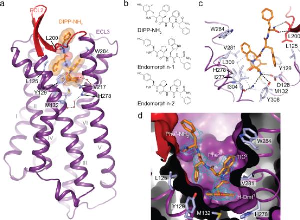
a, Overall view of δ-OR (purple cartoon, red ECL2) in complex with DIPP-NH2 (orange sticks and transparent spheres); residues lining the binding pocket are shown as light blue sticks, hydrogen bonds as black dotted lines, and water molecules as red spheres. b, Chemical structures of DIPP-NH2, endomorphin-1 and endomorphin-2 showing the structural similarities between the peptide analogue DIPP-NH2 and endogenous OR peptides. c, Close-up view of the DIPP-NH2 binding site; residues forming the DIPP-NH2 pocket are shown as light blue sticks. d, Sliced surface representation of the peptide binding pocket. The omit Fo-Fc electron density around the peptide DIPP-NH2 is contoured at 3σ and shown as a blue mesh.
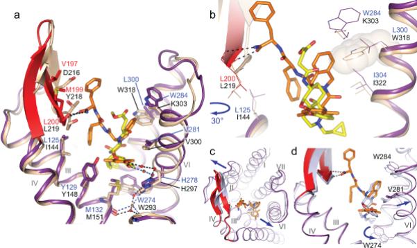
a, Superposition of the δ-OR structure (purple cartoon with red ECL2) bound to the bifunctional peptide DIPP-NH2 (orange sticks), and the μ-OR structure (beige cartoon) bound to β-FNA (yellow sticks). b, Superposition indicates that the Tic(2) group on DIPP-NH2 would clash with Trp318 (transparent beige sphere) on μ-OR. c, Superposition of BRILΔ36δ-OR–DIPP-NH2 (purple) and naltrindole-bound δ-OR (light blue) showing helix movements (indicated by arrows) observed upon DIPPNH2 binding. d, Close-up view of conformational changes occurring upon DIPP-NH2 binding compared to naltrindole bound receptor, including the shift of the Val2816.55 side chain. The change in orientation of the Trp2746.58 side chain in the naltrindole bound δ-OR structure is caused by the positioning of the cyclopentene group of naltrindole deeper into the receptor core.
Comment in
-
Serial femtosecond crystallography datasets from G protein-coupled receptors.Sci Data. 2016 Aug 1;3:160057. doi: 10.1038/sdata.2016.57. Sci Data. 2016. PMID: 27479354 Free PMC article.
References
Publication types
MeSH terms
Substances
Associated data
- Actions
- Actions
Grants and funding
- Y1-GM-1104/GM/NIGMS NIH HHS/United States
- U54 GM094599/GM/NIGMS NIH HHS/United States
- R01 GM095583/GM/NIGMS NIH HHS/United States
- R01 DA017204/DA/NIDA NIH HHS/United States
- U54 GM094618/GM/NIGMS NIH HHS/United States
- Y1-CO-1020/CO/NCI NIH HHS/United States
- R01 GM108635/GM/NIGMS NIH HHS/United States
- R37 DA004443/DA/NIDA NIH HHS/United States
- P01 DA035764/DA/NIDA NIH HHS/United States
- P41 GM103393/GM/NIGMS NIH HHS/United States
- R21 DA038858/DA/NIDA NIH HHS/United States
- R01 DA004443/DA/NIDA NIH HHS/United States
- U19 MH082441/MH/NIMH NIH HHS/United States
- R33 DA038858/DA/NIDA NIH HHS/United States
- MOP-89716/CAPMC/ CIHR/Canada
- DA-004443/DA/NIDA NIH HHS/United States
LinkOut - more resources
Full Text Sources
Other Literature Sources
Molecular Biology Databases
Research Materials
