. 2015 Mar 9;16(3):1050-61.
doi: 10.1021/acs.biomac.5b00099. Epub 2015 Feb 25.

Biomineralized matrices dominate soluble cues to direct osteogenic differentiation of human mesenchymal stem cells through adenosine signaling

Affiliations

Biomineralized matrices dominate soluble cues to direct osteogenic differentiation of human mesenchymal stem cells through adenosine signaling

Heemin Kang et al. Biomacromolecules. .

Abstract

Stem cell differentiation is determined by a repertoire of signals from its microenvironment, which includes the extracellular matrix (ECM) and soluble cues. The ability of mesenchymal stem cells (MSCs), a common precursor for the skeletal system, to differentiate into osteoblasts and adipocytes in response to their local cues plays an important role in skeletal tissue regeneration and homeostasis. In this study, we investigated whether a bone-specific calcium phosphate (CaP) mineral environment could induce osteogenic differentiation of human MSCs, while inhibiting their adipogenic differentiation, in the presence of adipogenic-inducing medium. We also examined the mechanism through which the mineralized matrix suppresses adipogenesis of hMSCs to promote their osteogenic differentiation. Our results show that hMSCs cultured on mineralized matrices underwent osteogenic differentiation despite being cultured in the presence of adipogenic medium, which indicates the dominance of matrix-based cues of the mineralized matrix in directing osteogenic commitment of stem cells. Furthermore, the mineralized matrix-driven attenuation of adipogenesis was reversed with the inhibition of A2b adenosine receptor (A2bR), implicating a role of adenosine signaling in mineralized environment-mediated inhibition of adipogenesis. Such synthetic matrices with an intrinsic ability to direct differentiation of multipotent adult stem cells toward a targeted phenotype while inhibiting their differentiation into other lineages not only will be a powerful tool in delineating the role of complex microenvironmental cues on stem cell commitment but also will contribute to functional tissue engineering and their translational applications.


Conflict of interest statement

Notes

The authors declare no competing financial interest.

Figures
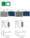
Figure 1
Figure 1
Characterization of biomineralized matrices. (a) Gross images of nonmineralized (NM) and mineralized (M) hydrogel discs. Scale bars represent 2 mm. (b) Scanning electron microscopy (SEM) images and corresponding energy dispersive spectra (EDS) of nonmineralized and mineralized matrices. Scale bars indicate 2 μm. Inset shows high-magnification images, and scale bar represents 500 nm. (c) Ca2+ and (d) PO43− amounts of nonmineralized and mineralized matrices after normalization to the dry weight of matrices. Release of (e) Ca2+ and (f) PO43− from mineralized matrices in Tris buffer lacking such ions at 37 °C as a function of time. Data are presented as the mean ± standard deviation (n = 3). Two groups were compared by employing two-tailed Student’s t-test. Asterisks were assigned to p-values with statistical significance (***, p < 0.001).
Figure 2
Figure 2
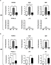
Quantitative PCR analyses of hMSCs cultured on various matrices under different medium conditions. Gene expressions of hMSCs for osteogenic markers (RUNX2, OCN, and BSP) as well as adipogenic markers (PPAR-γ2, αP2, and LPL) after 14 days of culture. Cells were cultured on nonmineralized (NM) and mineralized (M) matrices and coverslips (CS) in (a) growth medium (GM) and (b) adipogenic medium (AM). N.D. indicates a nondetectable amplification signal. Data are presented as the mean ± standard error (n = 3). Groups with different matrices in the same medium were compared by using one-way ANOVA with a Tukey-Kramer posthoc test. Asterisks were assigned to p-values with statistical significance (*, p < 0.05; **, p < 0.01; ***, p < 0.001).
Figure 3
Figure 3
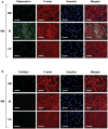
Immunofluorescent staining of (a) osteocalcin (green) and (b) perilipin (green) with corresponding F-actin (red) and nuclei (blue; Hoechst) for hMSCs on nonmineralized (NM) and mineralized (M) matrices and coverslips (CS) in growth medium (GM) after 14 days of culture. Scale bars represent 100 μm.
Figure 4
Figure 4
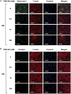
Immunofluorescent staining of (a) osteocalcin (green) and (b) perilipin (green) with corresponding F-actin (red) and nuclei (blue; Hoechst) for hMSCs on nonmineralized (NM) and mineralized (M) matrices and coverslips (CS) in adipogenic medium (AM) after 14 days of culture. Scale bars represent 100 μm.
Figure 5
Figure 5
Quantitative PCR analyses of hMSCs cultured on mineralized matrices under different medium conditions with varying amounts of A2bR antagonist, PSB 603. Fold expressions of hMSCs for osteogenic markers (RUNX2, OCN, and BSP) as well as adipogenic markers (PPAR-γ2, αP2, and LPL) after 14 days of culture. Cells were cultured in (a) growth medium (GM) and (b) adipogenic medium (AM) supplemented with PSB 603 at varying concentrations of 0, 0.5, 10, and 100 nM. N.D. indicates a nondetectable signal from PCR cycles. Data are presented as the mean ± standard error (n = 3). Groups with varying concentrations of PSB 603 under the same medium conditions were compared by using one-way ANOVA with a Tukey-Kramer posthoc test. Asterisks were assigned to p-values with statistical significance (*, p < 0.05; **, p < 0.01; ***, p < 0.001).
Figure 6
Figure 6
Immunofluorescent staining of (a) osteocalcin (green) and (b) perilipin (green) with corresponding F-actin (red) and nuclei (blue; Hoechst) for hMSCs on mineralized matrices after 14 days of culture in growth medium (GM) containing varying amounts (0, 0.5, 10, and 100 nM) of an A2bR antagonist, PSB 603. Scale bars represent 100 μm.
Figure 7
Figure 7
Immunofluorescent staining of (a) osteocalcin (green) and (b) perilipin (green) with corresponding F-actin (red) and nuclei (blue; Hoechst) for hMSCs on mineralized matrices after 14 days of culture in adipogenic medium (AM) containing varying amounts (0, 0.5, 10, and 100 nM) of an A2bR antagonist, PSB 603. Scale bars represent 100 μm.

Similar articles

Cited by

References

    1. Deans RJ, Moseley AB. Mesenchymal stem cells: biology and potential clinical uses. Exp Hematol. 2000;28:875–884. - PubMed
    1. Hwang NS, Zhang C, Hwang YS, Varghese S. Mesenchymal stem cell differentiation and roles in regenerative medicine. Wiley Interdiscip Rev: Syst Biol Med. 2009;1:97–106. - PubMed
    1. Baksh D, Song L, Tuan R. Adult mesenchymal stem cells: characterization, differentiation, and application in cell and gene therapy. J Cell Mol Med. 2004;8:301–316. - PMC - PubMed
    1. Beresford J, Bennett J, Devlin C, Leboy P, Owen M. Evidence for an inverse relationship between the differentiation of adipocytic and osteogenic cells in rat marrow stromal cell cultures. J Cell Sci. 1992;102:341–351. - PubMed
    1. Jaiswal RK, Jaiswal N, Bruder SP, Mbalaviele G, Marshak DR, Pittenger MF. Adult human mesenchymal stem cell differentiation to the osteogenic or adipogenic lineage is regulated by mitogen-activated protein kinase. J Biol Chem. 2000;275:9645–9652. - PubMed

Publication types

LinkOut - more resources