

Compounds targeting disulfide bond forming enzyme DsbB of Gram-negative bacteria
- PMID: 25686372
- PMCID: PMC4366281
- DOI: 10.1038/nchembio.1752
Compounds targeting disulfide bond forming enzyme DsbB of Gram-negative bacteria
Abstract
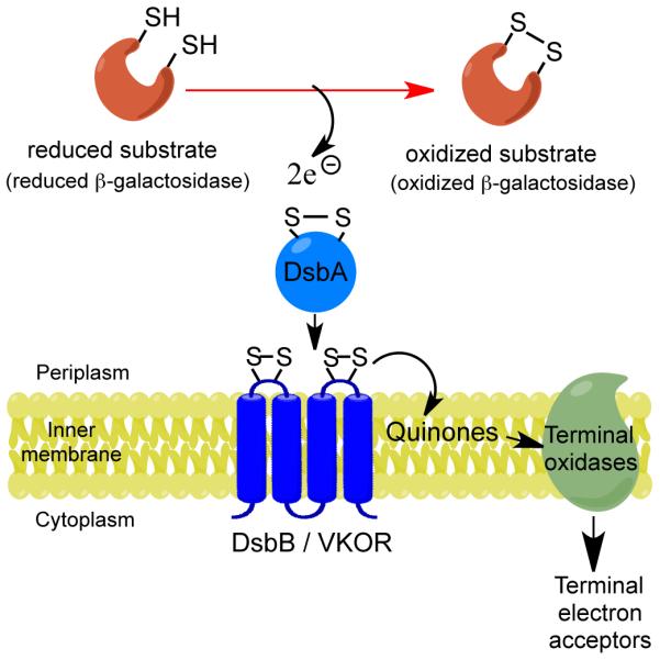
In bacteria, disulfide bonds confer stability on many proteins exported to the cell envelope or beyond. These proteins include numerous bacterial virulence factors, and thus bacterial enzymes that promote disulfide bond formation represent targets for compounds inhibiting bacterial virulence. Here, we describe a new target- and cell-based screening methodology for identifying compounds that inhibit the disulfide bond-forming enzymes Escherichia coli DsbB (EcDsbB) or Mycobacterium tuberculosis VKOR (MtbVKOR), which can replace EcDsbB, although the two are not homologs. Initial screening of 51,487 compounds yielded six specifically inhibiting EcDsbB. These compounds share a structural motif and do not inhibit MtbVKOR. A medicinal chemistry approach led us to select related compounds, some of which are much more effective DsbB inhibitors than those found in the screen. These compounds inhibit purified DsbB and prevent anaerobic growth of E. coli. Furthermore, these compounds inhibit all but one of the DsbBs of nine other Gram-negative pathogenic bacteria tested.
Figures


References
-
- Heras B, et al. DSB proteins and bacterial pathogenicity. Nat Rev Microbiol. 2009;7:215–25. - PubMed
-
- Depuydt M, Messens J, Collet JF. How proteins form disulfide bonds. Antioxid Redox Signal. 2011;15:49–66. - PubMed
-
- Bardwell JC, McGovern K, Beckwith J. Identification of a protein required for disulfide bond formation in vivo. Cell. 1991;67:581–9. - PubMed
Publication types
MeSH terms
Substances
Associated data
- PubChem-Substance/223733361
- PubChem-Substance/223733362
- PubChem-Substance/223733363
- PubChem-Substance/223733364
- PubChem-Substance/223733365
- PubChem-Substance/223733366
- PubChem-Substance/223733367
- PubChem-Substance/223733368
- PubChem-Substance/223733369
- PubChem-Substance/223733370
- PubChem-Substance/223733371
- PubChem-Substance/223733372
- PubChem-Substance/223733373
- PubChem-Substance/223733374
- PubChem-Substance/223733375
- PubChem-Substance/223733376
- PubChem-Substance/223733377
- PubChem-Substance/223733378
- PubChem-Substance/223733379
- PubChem-Substance/223733380
- PubChem-Substance/223733381
- PubChem-Substance/223733382
- PubChem-Substance/223733383
- PubChem-Substance/223733384
- PubChem-Substance/223733385
- PubChem-Substance/223733386
- PubChem-Substance/223733387
- PubChem-Substance/223733388
- PubChem-Substance/223733389
- PubChem-Substance/223733390
- PubChem-Substance/223733391
- PubChem-Substance/223733392
- PubChem-Substance/223733393
- PubChem-Substance/223733394
- PubChem-Substance/223733395
Grants and funding
- P01 HL087203/HL/NHLBI NIH HHS/United States
- P01 AI074805/AI/NIAID NIH HHS/United States
- 3P01-AI074805-04S1/AI/NIAID NIH HHS/United States
- GM041883/GM/NIGMS NIH HHS/United States
- R01 GM041883/GM/NIGMS NIH HHS/United States
- U54-AI057159/AI/NIAID NIH HHS/United States
- T32 AI007638/AI/NIAID NIH HHS/United States
- R01 HL092125/HL/NHLBI NIH HHS/United States
- UL1 RR025758/RR/NCRR NIH HHS/United States
- T32-HL07917/HL/NHLBI NIH HHS/United States
- R01-HL092125/HL/NHLBI NIH HHS/United States
- 5-UL1RR02568-01/RR/NCRR NIH HHS/United States
- U54 AI057159/AI/NIAID NIH HHS/United States
- P01-HL087203/HL/NHLBI NIH HHS/United States
- U54 HL112302/HL/NHLBI NIH HHS/United States
- T32 HL007917/HL/NHLBI NIH HHS/United States
LinkOut - more resources
Full Text Sources
Other Literature Sources
