. 2015 May:18:30-9.
doi: 10.1016/j.actbio.2015.02.005. Epub 2015 Feb 14.

Highly elastic and suturable electrospun poly(glycerol sebacate) fibrous scaffolds

Affiliations

Highly elastic and suturable electrospun poly(glycerol sebacate) fibrous scaffolds

Eric M Jeffries et al. Acta Biomater. 2015 May.

Abstract

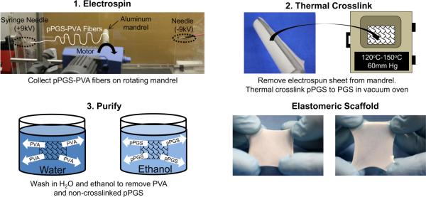
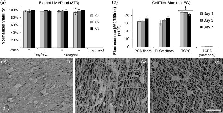
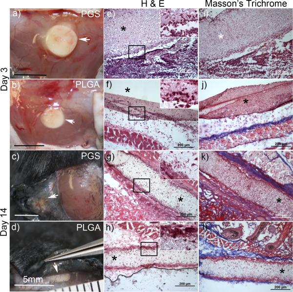
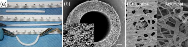
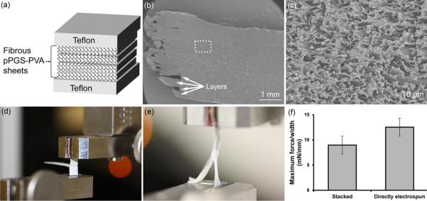
Poly(glycerol sebacate) (PGS) is a thermally-crosslinked elastomer suitable for tissue regeneration due to its elasticity, degradability, and pro-regenerative inflammatory response. Pores in PGS scaffolds are typically introduced by porogen leaching, which compromises strength. Methods for producing fibrous PGS scaffolds are very limited. Electrospinning is the most widely used method for laboratory scale production of fibrous scaffolds. Electrospinning PGS by itself is challenging, necessitating a carrier polymer which can affect material properties if not removed. We report a simple electrospinning method to produce distinct PGS fibers while maintaining the desired mechanical and cytocompatibility properties of thermally crosslinked PGS. Fibrous PGS demonstrated 5 times higher tensile strength and increased suture retention compared to porous PGS foams. Additionally, similar modulus and elastic recovery were observed. A final advantage of fibrous PGS sheets is the ability to create multi-laminate constructs due to fiber bonding that occurs during thermal crosslinking. Taken together, these highly elastic fibrous PGS scaffolds will enable new approaches in tissue engineering and regenerative medicine.

Keywords: Elastomer; Electrospinning; Fiber; Poly(glycerol sebacate) (PGS); Poly(vinyl alcohol) (PVA).


Figures

Figure 1
Figure 1
Fabrication of fibrous PGS sheets. 1. Electrospin pPGS-PVA to rotating (100rpm) mandrel 30cm from syringe needle at (+9kV), and 30cm from negatively- charged needle (−9kV). 2. Crosslink the sheet with high temperate and vacuum. 3. Purify samples by washing in water for 24h to remove soluble PVA. Rinse in graded ethanol dilutions to remove non-crosslinked pPGS.
Figure 2
Figure 2
SEM of PGS-PVA (55:45) fibers after electrospinning, thermal crosslinking, and purification. (a-b) Electrospun pPGS-PVA fibers. (c-e) PGS-PVA after crosslinking at respective conditions. (f-h) PGS-PVA fibers after washing in water for 24h. (i-k) PGS-PVA after ethanol purification. Inset: high magnification images of fibers. (*) Corresponding samples (d, e, j, k) after autoclaving. (Scale bar: 10μm for a, c-k and *; 5μm for all insets)
Figure 3
Figure 3
Purification analysis. (a) Percent of unwashed mass and thickness remaining after water and ethanol washes for each crosslinking conditions. (b) ATR-FTIR spectra of PVA, PGS and PGS-PVA during purification. (c) Heating and cooling curves from DSC measurements. Colors use the same legend as (b).
Figure 4
Figure 4
Mechanical testing. (a) Tensile testing plot of samples strained parallel (||) or perpendicular ( ⊥ ) to fiber axis. (b) Comparison of fibrous PGS to previously reported mechanical properties for porous and non-porous films under similar conditions. Note: UTS values for nonporous and porous films was during testing of dry samples. C2 crosslinking was 120°C for all samples. C5 crosslinking for porous and nonporous films was 150°C-48h but 120°C-24h, 150°C-24h for fibers. (c) Multi-cycle testing of C5 sample, strained 10-100% for 100 cycles. (d) Suture retention strength.
Figure 5
Figure 5
Cytocompatibility of PGS-PVA fibers. (a) Normalized viability of 3T3 cells cultured with extract from PGS-PVA samples. (b) CellTiter-Blue cell viability colorimetric assay with hcbEC. Metabolic activity of cells was measured by fluorescence emission at 590nm after 560nm excitation. All groups were significantly different from TCPS controls (asterisk). PGS fibers were not significantly different from PLGA fibers. (c) SEM of 3T3 cells on PGS-PVA fibers. Scale: 100 μm
Figure 6
Figure 6
Gross and histological images of fibrous PGS and PLGA implants after 3 and 14 days. Implants are indicated by arrowheads in gross images (a-d) of the subcutaneous tissue. Cross-sectional images stained by (H&E) are shown in (e-h) and Masson's trichrome (i-l) with implants indicated by an asterisk.
Figure 7
Figure 7
PGS-PVA fibrous conduits. (a) Small-diameter conduits demonstrate elastic properties and easy handling. (b) The fibrous morphology of these conduits can be observed in the SEM cross-section (Scale: 200 μm, Insert: 20 μm). (c) SEM images of the luminal and abluminal surfaces of the conduit reveal fibers with some fusion around the mandrel. (Scale: 10 μm)
Figure 8
Figure 8
Multi-laminate scaffolds. (a) Fibrous pPGS-PVA sheets were stacked between Teflon blocks and then thermally crosslinked. (b) SEM of purified multi-laminate structures reveals thick scaffolds with indistinguishable layers and (c) fibrous microstructure. (d) Image of dry laminated PGS sample in MTS grips before T-peel test. (e) Image of hydrated laminated PGS sample undergoing tensile T-peel testing. The free ends of the sample elongate while the two layers remain bonded together. (f) T-peel test maximum force/thickness before failing. Stacked samples were electrospun separately and laminated by stacking during crosslinking. In contrast, for directly electrospun samples, the second layer was electrospun onto the first layer.

Similar articles

Cited by

References

    1. Engler AJ, Sen S, Sweeney HL, Discher DE. Matrix elasticity directs stem cell lineage specification. Cell. 2006;126(4):677–89. - PubMed
    1. Discher DE, Janmey P, Wang YL. Tissue cells feel and respond to the stiffness of their substrate. Science. 2005;310(5751):1139–43. - PubMed
    1. Sabir MI, Xu X, Li L. A review on biodegradable polymeric materials for bone tissue engineering applications. Journal of Material Science. 2009;44:5713–24.
    1. Li Y, Thouas GA, Chen Q-Z. Biodegradable soft elastomers: synthesis/properties of materials and fabrication of scaffolds. Royal Society of Chemistry. 2012;2:8229–42.
    1. Rai R, Tallawi M, Grigore A, Boccaccini AR. Synthesis, properties and biomedical applications of poly(glycerol sebacate) (PGS): A review. Progress in Polymer Science. 2012;37:1051–78.

Publication types