. 2015 Apr 23;520(7548):563-6.
doi: 10.1038/nature14147. Epub 2015 Feb 9.

ATG14 promotes membrane tethering and fusion of autophagosomes to endolysosomes

Affiliations

ATG14 promotes membrane tethering and fusion of autophagosomes to endolysosomes

Jiajie Diao et al. Nature. .

Abstract

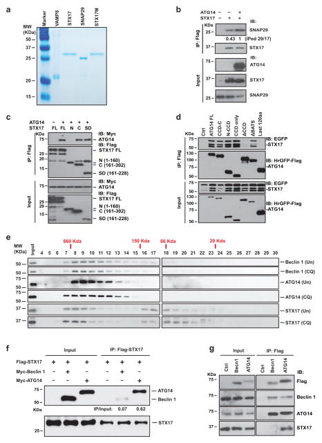
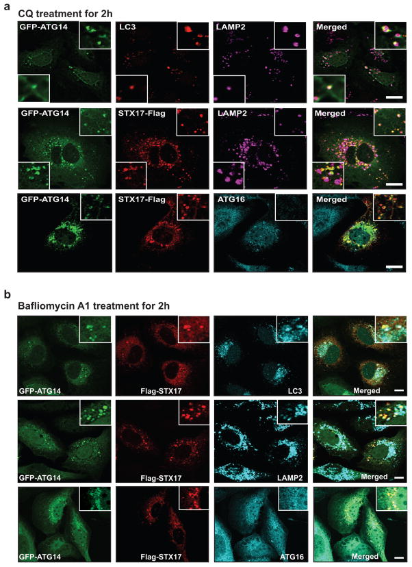
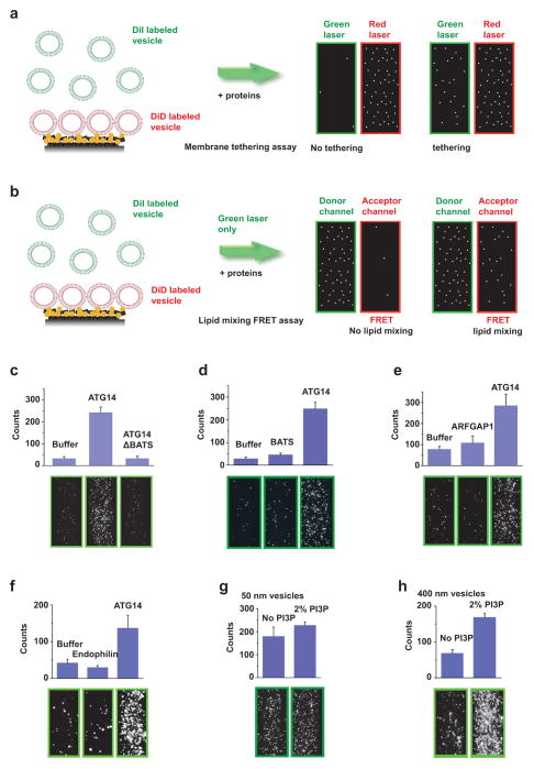
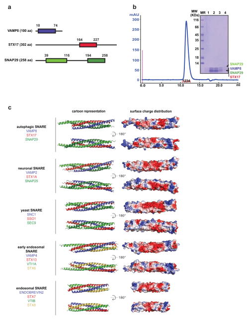
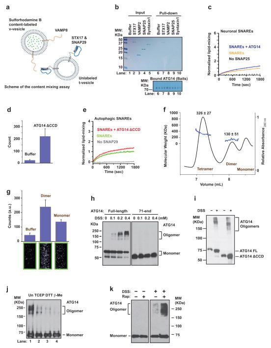
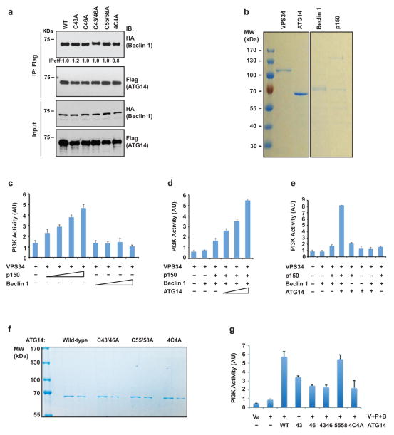
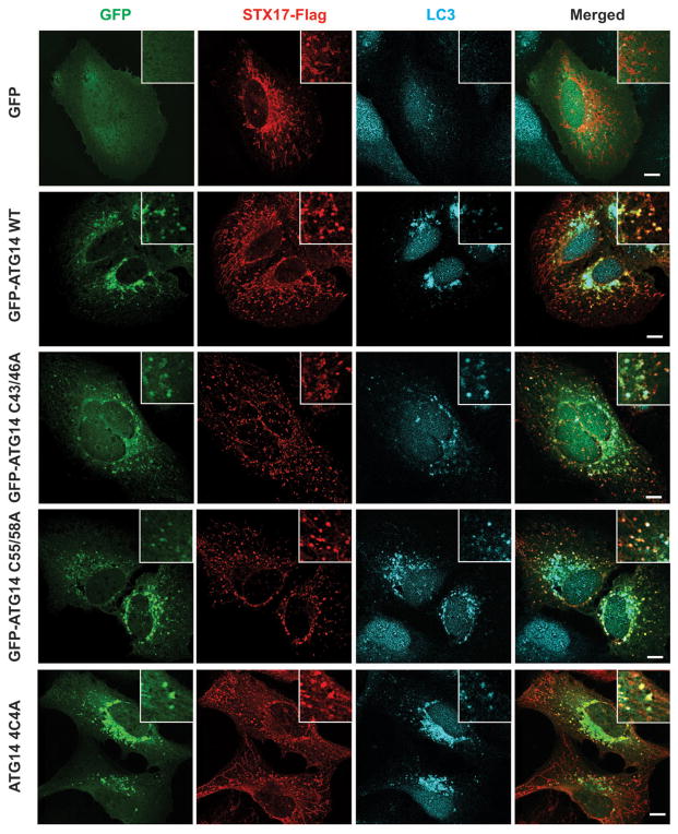
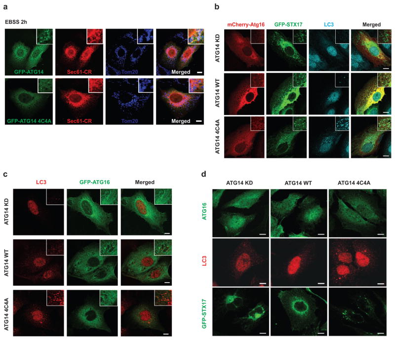
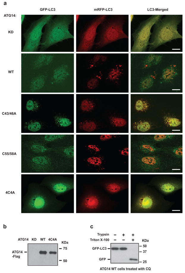
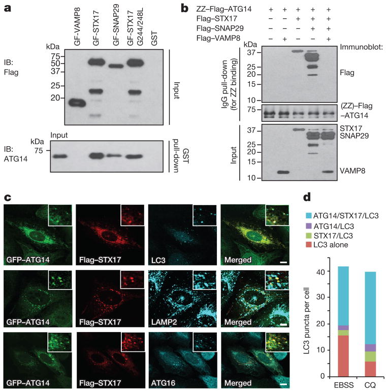
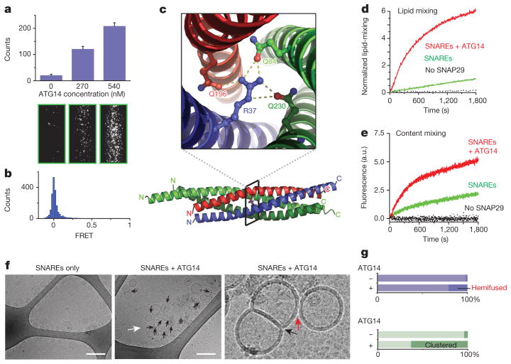
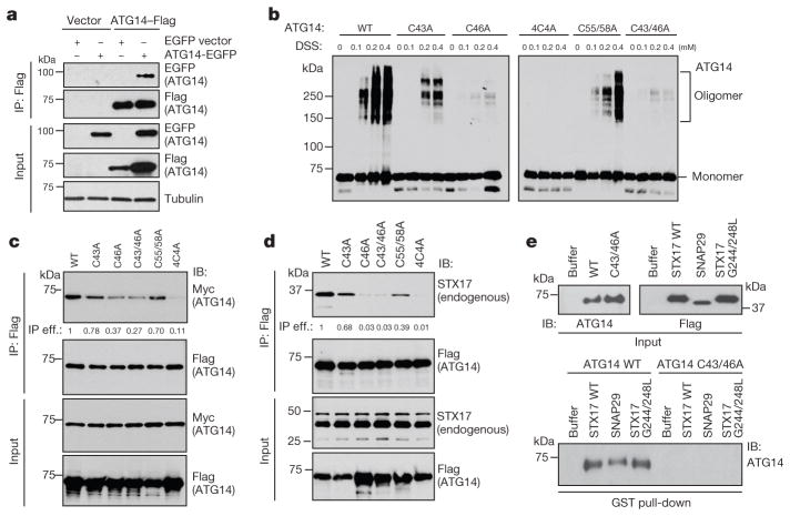
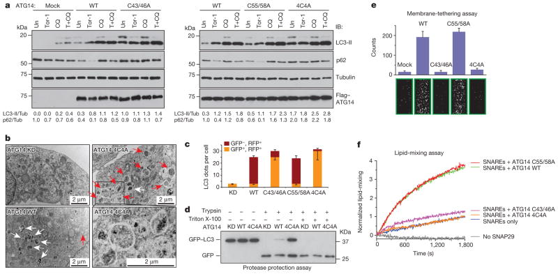
Autophagy, an important catabolic pathway implicated in a broad spectrum of human diseases, begins by forming double membrane autophagosomes that engulf cytosolic cargo and ends by fusing autophagosomes with lysosomes for degradation. Membrane fusion activity is required for early biogenesis of autophagosomes and late degradation in lysosomes. However, the key regulatory mechanisms of autophagic membrane tethering and fusion remain largely unknown. Here we report that ATG14 (also known as beclin-1-associated autophagy-related key regulator (Barkor) or ATG14L), an essential autophagy-specific regulator of the class III phosphatidylinositol 3-kinase complex, promotes membrane tethering of protein-free liposomes, and enhances hemifusion and full fusion of proteoliposomes reconstituted with the target (t)-SNAREs (soluble N-ethylmaleimide-sensitive factor attachment protein receptors) syntaxin 17 (STX17) and SNAP29, and the vesicle (v)-SNARE VAMP8 (vesicle-associated membrane protein 8). ATG14 binds to the SNARE core domain of STX17 through its coiled-coil domain, and stabilizes the STX17-SNAP29 binary t-SNARE complex on autophagosomes. The STX17 binding, membrane tethering and fusion-enhancing activities of ATG14 require its homo-oligomerization by cysteine repeats. In ATG14 homo-oligomerization-defective cells, autophagosomes still efficiently form but their fusion with endolysosomes is blocked. Recombinant ATG14 homo-oligomerization mutants also completely lose their ability to promote membrane tethering and to enhance SNARE-mediated fusion in vitro. Taken together, our data suggest an autophagy-specific membrane fusion mechanism in which oligomeric ATG14 directly binds to STX17-SNAP29 binary t-SNARE complex on autophagosomes and primes it for VAMP8 interaction to promote autophagosome-endolysosome fusion.


Conflict of interest statement

The authors declare no competing financial interests. Readers are welcome to comment on the online version of the paper.

Figures

Extended Data Figure 1
Extended Data Figure 1. Interaction among ATG14, STX17 and beclin 1
a, Coomassie-stained SDS gel of recombinantly expressed and purified full-length Flag-tagged STX17, SNAP29 and VAMP8. b, Overexpression of Myc-ATG14 stabilizes the STX17–SNAP29 binary t-SNARE complex in a co-immunoprecipitation assay. c, ATG14 interacts with the SNARE core domain of STX17 in a co-immunoprecipitation assay. d, The CCD domain of ATG14 interacts with STX17 in an co-immunoprecipitation assay. e, Fractionation of beclin 1, ATG14, STX17 with or without chloroquine treatment by Superdex 200. f, STX17 does not associate with beclin 1. Flag-tagged STX17 was co-transfected with either Myc-ATG14 or Myc-beclin 1 in HEK293T cells. ATG14 but not beclin 1 co-immunoprecipitated with STX17. The immunoprecipitation efficiency (immunoprecipitation/input) was normalized by the ratio of immunoprecipitated ATG14 or beclin 1 versus their inputs. g, STX17 interacts with ATG14 in a complex that is distinct from the beclin 1/PI3KC3 complex. Cell lysates from U2OS cells stably expressing Flag–beclin 1 or ATG14 were immunoprecipitated with anti-Flag M2 beads, and endogenous beclin 1, ATG14 and STX17 were detected in the immunoprecipitates.
Extended Data Figure 2
Extended Data Figure 2. Co-localization of ATG14 and STX17 on autophagosomes upon chloroquine or bafilomycin A1 treatments
a, b, GFP–ATG14 was co-transfected with STX17–Flag in chloroquine- (a) or bafilomycin-A1-treated (b) U2OS cells, and detected by GFP fluorescence and anti-Flag antibody in immunostaining. Endogenous LC3, LAMP1 or Atg16 were detected by anti-LC3, LAMP2 or Atg16 antibodies in immunostaining (n =20). Scale bars, 5 μm.
Extended Data Figure 3
Extended Data Figure 3. Characterization of ATG14-mediated membrane tethering
a, Scheme of the single-vesicle/liposome-tethering assay,. DiD-labelled liposomes were attached to the imaging surface through the interaction between biotin/NeutrAvidin; surface binding was assessed by red laser excitation (633 nm). Tethered DiI-labelled liposomes were detected by green laser excitation (532 nm). This assay was used in Figs 2a and 4e and Extended Data Figs 3c–h, 5d and 5g. b, Scheme of the FRET-based single-vesicle/liposome lipid-mixing assay. Tethered DiI-labelled liposomes were excited by illumination with a green laser (532 nm). Detection of emission in both green and red spectral regions was performed simultaneously by using a dichroic beam-splitter. The total number of tethered DiI-labelled liposomes were counted in the green fluorescence channel, and FRET to the DiD acceptor dyes was observed in the red fluorescence channel. This assay was only used in Fig. 2b. The field of view is 45 μm ×90 μm. See Methods for more details. c, The BATS domain deletion mutant of ATG14 (900 nM) does not promote liposome tethering. Top panels: quantitation. Bottom panels: representative fluorescence images of tethered liposomes (n =15). d, The BATS domain alone does not promote liposome tethering. Top panels: quantitation. Bottom panels: representative fluorescence images of tethered liposomes (n =15). e, Purified recombinant ADP-ribosylation factor GTPase-activating protein 1 (ARFGAP1, 870 nM) does not promote liposome tethering. Top panels: quantitation. Bottom panels: representative fluorescence images of tethered liposomes (n =15). f, Purified recombinant endophilin 1/Bif 1 (2 μM) does not promote liposome tethering. Top panels: quantitation. Bottom panels: representative fluorescence images of tethered liposomes (n =15). g, Incorporation of 2% PI3P into small liposomes (with a 50 nm diameter) failed to enhance the liposome-tethering activity by ATG14. Top panels: quantitation. Bottom panels: representative fluorescence images of tethered liposomes (n =15). h, Incorporation of 2% PI3P into large liposomes (with a 400 nm diameter) enhances the liposome-tethering activity by ATG14. Top panels: quantitation. Bottom panels: representative fluorescence images of tethered liposomes (n =15). Results in ch are presented as the mean (±s.d.) of random imaging locations (n =15) in the same sample channel.
Extended Data Figure 4
Extended Data Figure 4. Structure of the autophagic SNARE complex
a, Boundaries of SNARE domains in VAMP8, STX17 and SNAP29. b, Purification of the autophagic SNARE complex. The four SNARE core domains of VAMP8, STX17 and SNAP29 (with two SNARE core domains) were cloned, expressed in E. coli and co-purified (see Methods). The four fragments form a complex that appears as a single peak by SEC. The SDS–PAGE gel of the indicated four eluted fractions shows individual bands corresponding to the four SNARE core domains. Note that VAMP8 (10–74), STX17 (164–277) and SNAP29 (194–258) migrate at approximately the same position. The leftmost lane is a protein ladder with the molecular masses labelled. c, Comparison of SNARE structures. Shown are cartoon representation and surface charge distribution of the autophagic, neuronal, yeast, early endosomal and endosomal SNARE complex structures in two different orientations. The structures were placed such that the carboxy termini of the SNARE complexes are facing to the right. The surface charge distribution was generated using default ‘vacuum electrostatics’ in PyMOL for qualitative
Extended Data Figure 5
Extended Data Figure 5. Determining the specificity of ATG14 to autophagic SNAREs-mediated membrane fusion and characterizing recombinant ATG14 homo-oligomerization
a, Scheme of the ensemble content-mixing assay using autophagic SNARE-reconstituted proteoliposomes (v- and t-proteoliposomes). b, Purified recombinant Flag–STX17, His-VAMP2, His-SNAP25 and His-Syntaxin1 were incubated with IgG Sepharose associated with recombinant ZZ–Flag–ATG14, and the ATG14 binding proteins were cleaved by TEV protease and detected by Coomassie blue staining (upper panel). ZZ–Flag–ATG14 bound to IgG Sepahrose was detected by western blotting in the bottom panel. ATG14 binds to STX17 but not to neuronal SNAREs. c, ATG14 had no detectable effect on promoting ensemble lipid-mixing of proteoliposomes reconstituted with neuronal SNAREs (n =3). d, The membrane-tethering activity of ATG14 CCD deletion mutant is largely intact. Shown is the mean number of tethered vesicles (±s.d.) (n =15) in the same sample channel. e, The ATG14 CCD deletion mutant fails to enhance ensemble lipid mixing of proteoliposomes reconstituted with autophagic SNAREs (n =3). f, The oligomeric states of recombinant ATG14 were determined by SEC–MALS. g, Membrane-tethering activities of ATG14 monomer and dimer measured by the single protein-free vesicle/liposome membrane-tethering assay. Shown are the mean numbers of tethered vesicles (±s.d.) (n =15) in the same sample channel. Representative images are shown in the bottom panels (n =15). h, Mapping ATG14 oligomerization sites to its N terminus. Flag-tagged full-length ATG14 or an ATG14 truncating mutant (lacking the first 70 residues) were transfected into HEK293T cells and treated with cross-linking agent DSS (0, 0.1, 0.2, 0.4 mM) for 30 min, then subjected to SDS–PAGE analysis. ATG14 was probed by anti-Flag antibody. i, ATG14 CCD deletion mutant still forms oligomer in the DSS cross-linking assay. j, Purified recombinant ATG14 (36 nM) was boiled in non-reducing SDS sample buffer with 12.5 mM TCEP (lane 2), 25 mM DTT (lane 3) or 2.5% β-mercaptoethanol (lane 4), or mock-treated (lane 1), and the samples were loaded on non-reducing SDS–PAGE and probed for anti-Flag antibody for ATG14. k, Flag–ATG14 was transfected into HEK293T cells and treated with rapamycin (500 nM for 12 h) and/or cross-linking agent DSS (0.2 μM) for 30 min, then subjected to SDS–PAGE analysis. ATG14 was probed by anti-Flag antibody.
Extended Data Figure 6
Extended Data Figure 6. ATG14 homo-oligomerization is not essential for beclin 1 interaction and PI3KC3 activation
a, Interaction between Flag-tagged WT ATG14 and mutants with HA-tagged beclin 1 observed in a co-immunoprecipitation assay. b, Purification of Flag-tagged PI3KC3 complex subunits from insect cells. c, Graph showing p150 but not beclin 1 stimulated Vps34 lipid kinase activity using in vitro TLC kinase assays. Shown are the mean intensity values of radioactive PI3P spots (±s.d.) (n =3). AU, arbitrary unit. d, ATG14 enhanced the lipid kinase activity of Vps34–p150–beclin 1. Shown are the mean intensity values of radioactive PI3P spots (±s.d.) (n =3). e, The requirement of beclin 1 and p150 to the promotion effect of ATG14. Shown are the mean intensity values of radioactive PI3P spots (±s.d.) (n =3). f, Purification of ATG14 WT and mutants from insect cells. Two doses of each recombinant protein were loaded. g, Effect of ATG14 WT and mutants on Vps34(V)–p150(P)–beclin 1(B) lipid kinase activity. Shown are the mean intensity values of radioactive PI3P spots (±s.d.) (n =3).
Extended Data Figure 7
Autophagosome targeting of ATG14 homo-oligomerization-deficient mutants. GFP, GFP–ATG14 WT, GFP–ATG14 C43/46A, GFP–ATG14 C55/58A and GFP–ATG14 4C4A were co-transfected with STX17–Flag in U2OS cells and detected by immunostaining. Endogenous LC3 was detected by anti-LC3 antibody in immunostaining (n =20). Scale bars, 5 μm.
Extended Data Figure 8
Extended Data Figure 8. Characterization of the subcellular localization of ATG14 HOD mutants
a, U2OS cells transfected with GFP–ATG14 WT or ATG14 4C4A and mCherry-Sec61β (ER marker) were treated with EBSS for 2 h and co-stained with endogenous Tom20 (mitochondria marker). Both GFP–ATG14 and GFP–ATG14 4C4A were found at the junction of ER (Sec61β) and mitochondria (Tom20) (n =20). Scale bars, 5 μm. b, Accumulation of endogenous LC3 and GFP–STX17, but not mCherry-Atg16, in ATG14 4C4A mutant but not in ATG14 WT or knockdown (KD) cells (n =20). Scale bars, 5 μm. c, ATG14 knockdown U2OS cells stably complemented with ATG14 WT or 4C4A mutant ATG14 were transfected with GFP–ATG16, and endogenous LC3 staining and ATG16 fluorescence were imaged. LC3 but not ATG16 puncta were accumulated in ATG14 4C4A cells (n =20). Scale bars, 5 μm. d, ATG14 knockdown U2OS cells stably complemented with ATG14 WT or 4C4A mutant ATG14 were transfected with GFP–STX17, and endogenous ATG16 staining and STX17 fluorescence were imaged. STX17 but not ATG16 puncta were accumulated in ATG14 4C4A cells (n =20). Scale bars, 5 μm.
Extended Data Figure 9
Extended Data Figure 9. ATG14 homo-oligomerization is required for autophagosome maturation
a, mRFP–GFP–LC3, with WT or mutant ATG14, were expressed in U2OS cells, and LC3 puncta (overlapped green and red fluorescence) in ATG14-expressing cells were imaged (n =20). Scale bars, 5 μm. Yellow LC3 signals represent phagophores or autophagosomes before fusion. Red-only LC3 signals represent acidified mature autophagosomes. Un, untreated. Rap, rapamycin. The corresponding quantitative statistical analysis is shown in Fig. 4c. b, Stable expression of Flag–ATG14 or Flag–ATG14 4C4A in ATG14 knockdown U2OS cells. c, Protease protection assay in U2OS ATG14 WT cells treated with chloroquine (2 h). U2OS ATG14 WT cells were infected with lentivirus expressing GFP–LC3 for 24 h and treated with chloroquine 2 h before harvest. The autophagosome-enriched fraction from these cells was isolated by centrifugation, and incubated with trypsin (10 μg ml−1) with and without 0.4% Triton X-100 for 25 min at 30 °C. GFP or GFP–LC3 were detected by western blotting.
Figure 1
Figure 1. ATG14 interacts with STX17–SNAP29 on mature autophagosomes
a, Interaction between purified recombinant ATG14 and GST–Flag-tagged (GF) autophagic SNAREs using an in vitro GST pull-down assay followed by western blot (IB, immunoblot). b, Recombinant ATG14 binds to STX17–SNAP29 binary t-SNARE complex but not STX17–SNAP29–VAMP8 ternary complex using an in vitro immunoglobulin-G (IgG) pull-down assay followed by western blot. c, Flag–STX17, GFP–ATG14, endogenous LC3, LAMP2 and ATG16 were detected by immunostaining in EBSS-starved U2OS cells (n =20). Scale bars, 5 μm. d, Corresponding statistical analysis of co-localization of ATG14, STX17 and LC3 upon treatment with EBSS or chloroquine (CQ).
Figure 2
Figure 2. ATG14 promotes membrane tethering and enhances autophagic SNARE-mediated fusion
a, Purified recombinant ATG14 promotes protein-free single liposome tethering (Methods and Extended Data Fig. 3a). Top: mean number of tethered liposomes (±s.d.) (n =15) at random locations in the sample chamber; bottom: corresponding representative images (n =15). b, Fluorescence resonance energy transfer (FRET) efficiency profile between single donor/acceptor-dye liposome pairs upon addition of recombinant ATG14 (Methods and Extended Data Fig. 3b). c, The crystal structure of the autophagic SNARE complex is shown at the bottom and a close-up view of the ionic layer at the centre is shown at the top. d, ATG14 enhances ensemble lipid-mixing of proteoliposomes reconstituted with autophagic SNAREs (n =3). e, ATG14 enhances ensemble content-mixing of proteoliposomes reconstituted with autophagic SNAREs (n =3); a.u., arbitrary units. f, Representative cryo-electron micrographs (n =20) of proteoliposomes reconstituted with autophagic SNAREs. Left: without ATG14. Middle: with ATG14; black and white arrows indicate hemifusion diaphragms and proteoliposome clusters, respectively. Right: close-up view; black and red arrows indicate a hemifused diaphragm and a tethered proteoliposome pair, respectively. Scale bars, 200 nm. g, Percentage of hemifused or clustered proteoliposomes micrographs (n =20).
Figure 3
Figure 3. ATG14 homo-oligomerization is required for autophagic SNARE binding
a, Immunoprecipitation (IP) of ATG14–eGFP by ATG14–Flag transfected into HEK293T cells. b, Mapping ATG14 oligomerization sites to its cysteine repeats by cross-linking with DSS and analysed by SDS–polyacrylamide gel electrophoresis (SDS–PAGE). c, Interaction between Flag-tagged wild-type (WT) ATG14 and mutants with Myc-ATG14 observed in a co-immunoprecipitation assay. IP eff., immunoprecipitation efficiency. d, Interaction between Flag-tagged WT ATG14 and mutants with endogenous STX17 observed in a co-immunoprecipitation assay. The immunoprecipitation efficiency (immunoprecipitation/input) in c and d was normalized by the ratio of immunoprecipitated Flag–ATG14 or interactants versus their inputs. e, In vitro GST pull-down of purified recombinant WT ATG14 or C43A/C46A mutant with GST-tagged autophagic SNAREs followed by western blot.
Figure 4
Figure 4. ATG14 homo-oligomerization is required for autophagosomal fusion with endolysosomes in vivo and in vitro
a, Autophagic flux analysis in ATG14-deficient U2OS cells reconstituted with WT ATG14 and mutants. Un, untreated; Tor-1, Torin 1. b, Accumulation of complete double-membrane autophagosome (red arrows) under TEM in ATG14 4C4A mutant but not in ATG14 WT or knockdown (KD) cells treated with rapamycin (n =15). White arrows denote autolysosomes. c, Quantitative analysis of acidified autophagosomes (GFPRFP+) versus neutral autophagosomes (GFP+RFP+) per cell in rapamycin-treated ATG14 knockdown U2OS cells expressing WT or mutant ATG14 transfected with mRFP–GFP–LC3 (mean ±s.d.) (n =20). d, GFP–LC3 was protected from trypsin digestion in ATG14 4C4A mutant but not in ATG14 WT or knockdown cells. e, Tethering of protein-free liposomes by recombinant WT ATG14 and specified mutants using the single-vesicle membrane-tethering assay (mean number of tethered liposomes ±s.d.) (n =20)). f, Ensemble lipid-mixing activity of WT ATG14 and mutants between proteoliposomes reconstituted with autophagic SNAREs (n =3).

References

    1. Levine B, Klionsky DJ. Development by self-digestion: molecular mechanisms and biological functions of autophagy. Dev Cell. 2004;6:463–477. - PubMed
    1. Huang WP, Klionsky DJ. Autophagy in yeast: a review of the molecular machinery. Cell Struct Funct. 2002;27:409–420. - PubMed
    1. Moreau K, Ravikumar B, Renna M, Puri C, Rubinsztein DC. Autophagosome precursor maturation requires homotypic fusion. Cell. 2011;146:303–317. - PMC - PubMed
    1. Nair U, et al. SNARE proteins are required for macroautophagy. Cell. 2011;146:290–302. - PMC - PubMed
    1. Ragusa MJ, Stanley RE, Hurley JH. Architecture of the Atg17 complex as a scaffold for autophagosome biogenesis. Cell. 2012;151:1501–1512. - PMC - PubMed

Publication types

MeSH terms

Associated data