. 2015 Feb 17:5:8499.
doi: 10.1038/srep08499.

Static stretch affects neural stem cell differentiation in an extracellular matrix-dependent manner

Affiliations

Static stretch affects neural stem cell differentiation in an extracellular matrix-dependent manner

Janahan Arulmoli et al. Sci Rep. .

Abstract

Neural stem and progenitor cell (NSPC) fate is strongly influenced by mechanotransduction as modulation of substrate stiffness affects lineage choice. Other types of mechanical stimuli, such as stretch (tensile strain), occur during CNS development and trauma, but their consequences for NSPC differentiation have not been reported. We delivered a 10% static equibiaxial stretch to NSPCs and examined effects on differentiation. We found static stretch specifically impacts NSPC differentiation into oligodendrocytes, but not neurons or astrocytes, and this effect is dependent on particular extracellular matrix (ECM)-integrin linkages. Generation of oligodendrocytes from NSPCs was reduced on laminin, an outcome likely mediated by the α6 laminin-binding integrin, whereas similar effects were not observed for NSPCs on fibronectin. Our data demonstrate a direct role for tensile strain in dictating the lineage choice of NSPCs and indicate the dependence of this phenomenon on specific substrate materials, which should be taken into account for the design of biomaterials for NSPC transplantation.


Figures

Figure 1
Figure 1. Physical regulators of stem cell behavior.
Stem cells experience a variety of physical cues within the natural microenvironment that can have significant effects on survival, proliferation, differentiation, and gene expression. Examples of these mechanical cues include shear force from fluid, mechanical stress from cell-cell interactions and cell movements, and surface topology and substrate stiffness through components of the ECM and surrounding cells.
Figure 2
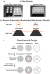
Figure 2. Induction of 10% equibiaxial static stretch to adhered NSPCs via the J-flex device.
(a) J-Flex device: white polytetrafluoroethylene (Teflon) disks (25 mm diameter) attached to black rubber corks (lower plate) press fit into a Flexcell Bioflex plate (standard 6-well size) with silicone elastomer membranes (upper plate). When the two plates are attached, a 10% equibiaxial strain is induced on the silicone elastomer membranes (attached plates). Rubber bands (not shown) were used to keep the plates firmly press-fit. (b) Device schematic illustrating membrane stretch: top and side views of the membrane (orange circles in top view; orange line in side view), cork (black) and Teflon disk (grey) when the plates are attached. The stretched configuration shows displacement (10% equibiaxial) of two markings on the membrane in the top view and the corresponding setup with the cork and disk in the side view (not to scale). (c) Experimental design: mNSPCs were seeded on laminin-coated surfaces (membranes and glass) for 18 hours in proliferation conditions, followed by application of mechanical stimulus (stretched group only) after removal of growth factors (differentiation conditions) to assess formation of neurons (3 days), astrocytes (7 days), or oligodendrocytes (5 or 7 days) by immunostaining post-differentiation.
Figure 3
Figure 3. Stretch inhibits mNSPC differentiation into oligodendrocytes.
(a) Mouse cortical NSPCs and (b) rat hippocampal NSPCs differentiated on unstretched and stretched membranes or glass were stained with oligodendrocyte cell surface marker O4 and Hoechst for nuclear DNA. Static stretch reduces oligodendrocyte differentiation regardless of cell source. (a) P = 0.0003 (unstretched vs. stretched), P = 0.002 (glass vs. stretched). (b) P = 0.001 (unstretched vs. stretched), P = 0.016 (glass vs. stretched). **P < 0.01, *P < 0.05. Error bars represent SEM. N = 3 independent biological repeats.
Figure 4
Figure 4. ECMs and integrins regulate mNSPC differentiation in response to static stretch.
(a) Cells in the oligodendrocyte lineage express the laminin-binding integrin α6β1 but are driven to proliferate by fibronectin via the αVβ3 integrin. (b) Delivery of a static stretch to mNSPCs on fibronectin does not affect differentiation into oligodendrocytes (no significant difference between unstretched vs. stretched group) although there is a difference between the stretched and glass groups. *P < 0.05. P = 0.013 (stretched vs. glass). Error bars represent SEM. N = 3 independent biological repeats. (c) Oligodendrocyte lineage cells can bind laminin through the α6β1 integrin to affect differentiation. (d) Inhibiting α6 integrin with a function-blocking antibody negates the effect of stretch on oligodendrocyte differentiation from mNSPCs as shown by a lack of decreased O4 expression for cells on stretched membranes compared to those on unstretched membranes treated with α6 integrin antibody. *P < 0.05, P = 0.013 (no antibody). P = 0.025 (IgG2A). Error bars represent SEM. N = 3 independent biological repeats.
Figure 5
Figure 5. Effects of stretched membrane stiffness on NSPC differentiation.
(a) Pre-stretched membranes were seeded with mNSPCs when the J-Flex device was already in place in order to control for the stiffness increase associated with the application of stretch at the onset of differentiation. Application of stretch to the membranes is denoted by red arrows. (b) Increase in stiffness upregulates oligodendrocyte differentiation as illustrated by the difference in percentage of O4-positive cells on pre-stretched vs. unstretched membranes. P = 0.0003 (unstretched vs. stretched). P = 0.01 (unstretched vs. pre-stretched). P = 1.9E−0.6 (pre-stretched vs. stretched). P = 0.002 (glass vs. stretched). P = 0.003 (glass vs. pre-stretched). (c) Stiffer membranes promote differentiation of more neurons from mNSPCs as demonstrated by significantly more MAP2-positive cells on pre-stretched or stretched membranes compared to cells on unstretched membranes. P = 0.002 (unstretched vs. stretched). P = 0.04 (unstretched vs. pre-stretched). (d) Negligible effects of stiffness or stretch on astrocyte generation as shown by minimal differences in GFAP-positive cells between stretched and pre-stretched against unstretched groups. P = 0.03 (pre-stretched vs. stretched). *P < 0.05, **P < 0.01, ***P < 0.0001. Error bars represent SEM. N = 3 independent biological repeats.

References

    1. Gage F. H. Mammalian neural stem cells. Science 287, 1433–1438 (2000). - PubMed
    1. Kshitiz. et al. Control of stem cell fate and function by engineering physical microenvironments. Integr Biol 4, 1008–1018 (2012). - PMC - PubMed
    1. Engler A., Sen S., Sweeney H. & Discher D. Matrix elasticity directs stem cell lineage specification. Cell 126, 677–689 (2006). - PubMed
    1. Tyler W. J. The mechanobiology of brain function. Nat Rev Neurosci 13, 867–878 (2012). - PubMed
    1. Saha K. et al. Substrate modulus directs neural stem cell behavior. Biophys J 95, 4426–4438 (2008). - PMC - PubMed

Publication types