

Multilayered Nanoparticles for Gene Delivery Used to Reprogram Human Foreskin Fibroblasts to Neurospheres
- PMID: 25687130
- PMCID: PMC4523045
- DOI: 10.1089/ten.TEC.2014.0482
Multilayered Nanoparticles for Gene Delivery Used to Reprogram Human Foreskin Fibroblasts to Neurospheres
Abstract
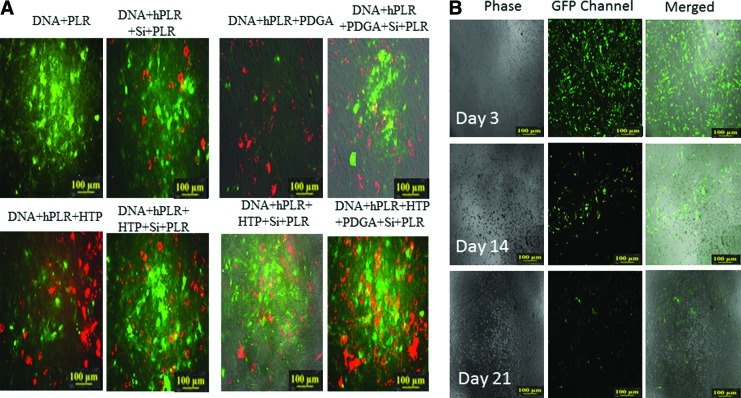
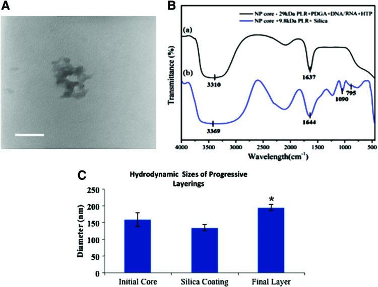
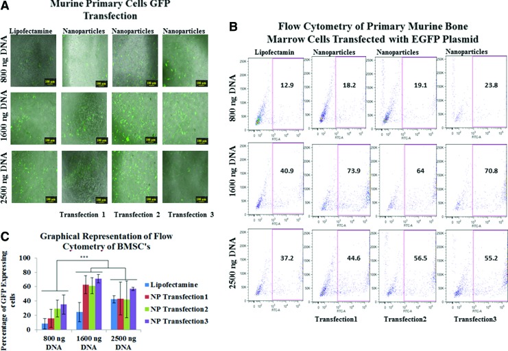
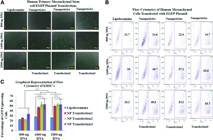
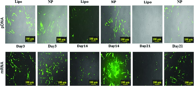
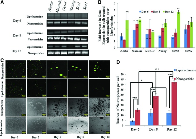
Polycationic nanocomplexes are a robust means for achieving nucleic acid condensation and efficient intracellular gene deliveries. To enhance delivery, a multilayered nanoparticle consisting of a core of electrostatically bound elements was used. These included a histone-mimetic peptides, poly-l-arginine and poly-d-glutamic acid was coated with silicate before surface functionalization with poly-l-arginine. Transfection efficiencies and duration of expression were similar when using green fluorescent protein (GFP) plasmid DNA (pDNA) or GFP mRNA. These nanoparticles demonstrated significantly higher (>100%) and significantly longer (15 vs. 4 days) transfection efficiencies in comparison to a commercial transfection agent (Lipofectamine 2000). Reprogramming of human foreskin fibroblasts using mRNA to the Sox2 transcription factor resulted in three-fold higher neurosphere formation in comparison to the commercial reagent. These results demonstrate the potential of these nanoparticles as ideal vectors for gene delivery.
Figures
Similar articles
-
Retro-inverso d-peptide-modified hyaluronic acid/bioreducible hyperbranched poly(amido amine)/pDNA core-shell ternary nanoparticles for the dual-targeted delivery of short hairpin RNA-encoding plasmids.Acta Biomater. 2017 Jul 15;57:156-169. doi: 10.1016/j.actbio.2017.04.024. Epub 2017 Apr 22. Acta Biomater. 2017. PMID: 28442415
-
Poly(2-hydroxyethyl methacrylate)-b-poly(L-Lysine) cationic hybrid materials for non-viral gene delivery in NIH 3T3 mouse embryonic fibroblasts.Macromol Biosci. 2014 Sep;14(9):1239-48. doi: 10.1002/mabi.201400071. Epub 2014 May 23. Macromol Biosci. 2014. PMID: 24862905
-
Polyethylenimine-based amphiphilic core-shell nanoparticles: study of gene delivery and intracellular trafficking.Biointerphases. 2012 Dec;7(1-4):16. doi: 10.1007/s13758-011-0016-4. Epub 2012 Feb 9. Biointerphases. 2012. PMID: 22589059
-
Polycationic nanoparticles as nonviral vectors employed for gene therapy in vivo.Mini Rev Med Chem. 2010 Feb;10(2):126-37. doi: 10.2174/138955710791185127. Mini Rev Med Chem. 2010. PMID: 20408797 Review.
-
Multifunctional nanocomplexes for gene transfer and gene therapy.Cell Biol Toxicol. 2010 Feb;26(1):69-81. doi: 10.1007/s10565-009-9141-y. Epub 2010 Feb 3. Cell Biol Toxicol. 2010. PMID: 20127400 Review.
Cited by
-
Effective gene delivery using size dependant nano core-shell in human cervical cancer cell lines by magnetofection.PLoS One. 2023 Sep 7;18(9):e0289731. doi: 10.1371/journal.pone.0289731. eCollection 2023. PLoS One. 2023. PMID: 37676882 Free PMC article.
-
Polymeric Amines and Ampholytes Derived from Poly(acryloyl chloride): Synthesis, Influence on Silicic Acid Condensation and Interaction with Nucleic Acid.Polymers (Basel). 2017 Nov 16;9(11):624. doi: 10.3390/polym9110624. Polymers (Basel). 2017. PMID: 30965927 Free PMC article.
References
-
- Musick M.A., McConnell K.I., Lue J.K., Wei F., Chen C., and Suh J. Reprogramming virus nanoparticles to bind metal ions upon activation with heat. Biomacromolecules 12, 2153, 2011 - PubMed
-
- Li H., Park S.H., Reif J.H., LaBean T.H., and Yan H. DNA-templated self-assembly of protein and nanoparticle linear arrays. J Am Chem Soc 126, 418, 2004 - PubMed
-
- Huangfu D., Osafune K., Maehr R., Guo W., Eijkelenboom A., Chen S., Muhlestein W., and Melton D.A. Induction of pluripotent stem cells from primary human fibroblasts with only Oct4 and Sox2. Nat Biotechnol 26, 1269, 2008 - PubMed
Publication types
MeSH terms
Substances
Grants and funding
LinkOut - more resources
Full Text Sources
Other Literature Sources
