

Induced Pluripotent Stem (iPS) Cell Culture Methods and Induction of Differentiation into Endothelial Cells
- PMID: 25687301
- PMCID: PMC4539286
- DOI: 10.1007/7651_2015_203
Induced Pluripotent Stem (iPS) Cell Culture Methods and Induction of Differentiation into Endothelial Cells
Abstract
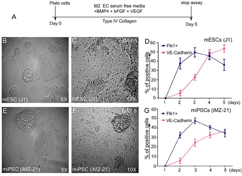
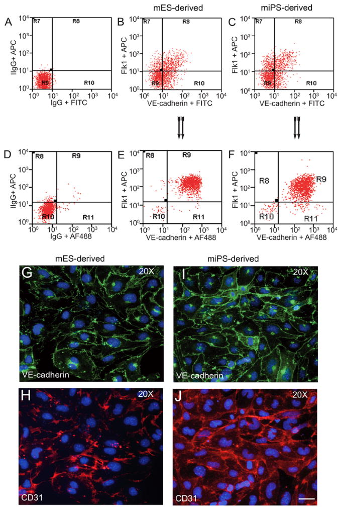
The study of stem cell behavior and differentiation in a developmental context is complex, time-consuming, and expensive, and for this reason, cell culture remains a method of choice for developmental and regenerative biology and mechanistic studies. Similar to ES cells, iPS cells have the ability to differentiate into endothelial cells (ECs), and the route for differentiation appears to mimic the developmental process that occurs during the formation of an embryo. Traditional EC induction methods from embryonic stem (ES) cells rely mostly on the formation of embryoid body (EB), which employs feeder or feeder-free conditions in the presence or absence of supporting cells. Similar to ES cells, iPS cells can be cultured in feeder layer or feeder-free conditions. Here, we describe the iPS cell culture methods and induction differentiation of these cells into ECs. We use anti-mouse Flk1 and anti-mouse VE-cadherin to isolate and characterize mouse ECs, because these antibodies are commercially available and their use has been described in the literature, including by our group. The ECs produced by this method have been used by our laboratory, and we have demonstrated their in vivo potential. We also discuss how iPS cells differ in their ability to differentiate into endothelial cells in culture.
Keywords: Angiogenesis; CD31; Endothelial cells; Flk1; Klf4; Nanog; Oct4; Sox2; VE-cadherin; iPS cells.
Figures
References
-
- Evans MJ, Kaufman MH. Establishment in culture of pluripotential cells from mouse embryos. Nature. 1981;292:154–156. - PubMed
-
- Bradley A, Robertson E. Embryo-derived stem cells: a tool for elucidating the developmental genetics of the mouse. Curr Top Dev Biol. 1986;20:357–371. - PubMed
-
- Nagy A, Gócza E, Diaz EM, Prideaux VR, Iványi E, Markkula M, Rossant J. Embryonic stem cells alone are able to support fetal development in the mouse. Development. 1990;110(3):815–821. - PubMed
-
- Wilmut I, Schnieke AE, McWhir J, Kind AJ, Campbell KH. Viable offspring derived from fetal and adult mammalian cells. Nature. 1997;385:810–813. - PubMed
Publication types
MeSH terms
Substances
Grants and funding
LinkOut - more resources
Full Text Sources
Other Literature Sources
Research Materials
