. 2015 Oct;240(10):1298-309.
doi: 10.1177/1535370215570191. Epub 2015 Feb 15.

Temporal responses of human endothelial and smooth muscle cells exposed to uniaxial cyclic tensile strain

Affiliations

Temporal responses of human endothelial and smooth muscle cells exposed to uniaxial cyclic tensile strain

Alexandra M Greiner et al. Exp Biol Med (Maywood). 2015 Oct.

Abstract

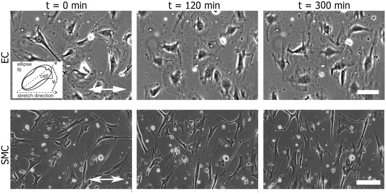
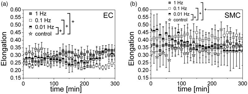
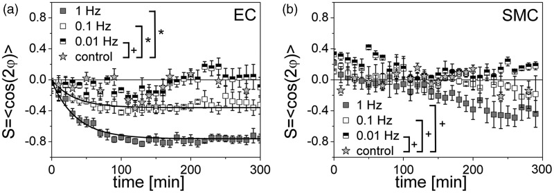
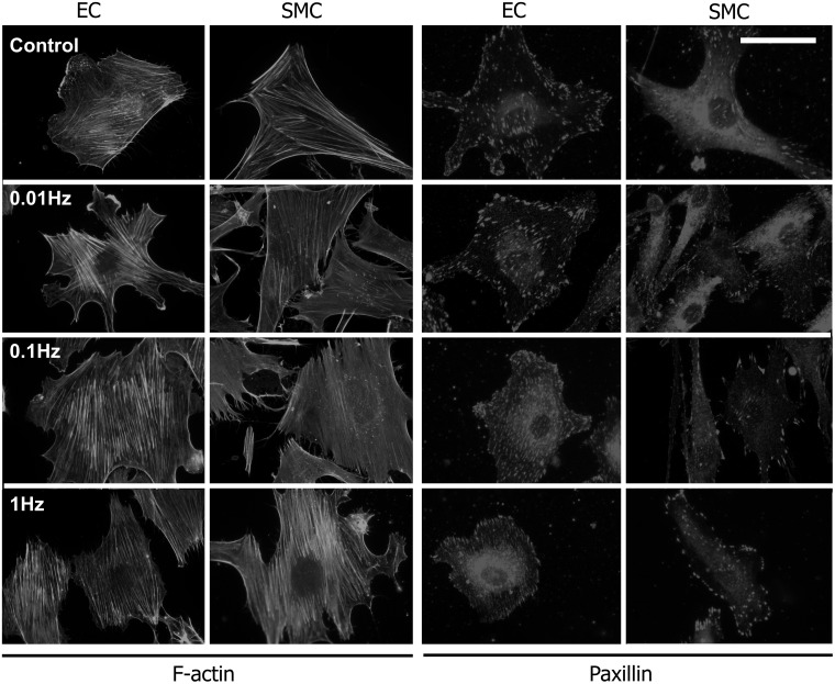
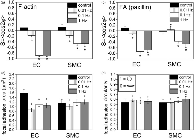
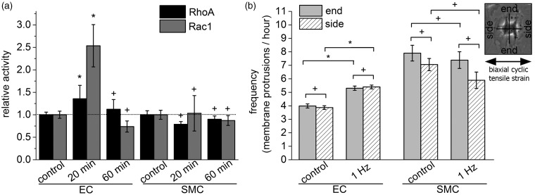
The physiology of vascular cells depends on stimulating mechanical forces caused by pulsatile flow. Thus, mechano-transduction processes and responses of primary human endothelial cells (ECs) and smooth muscle cells (SMCs) have been studied to reveal cell-type specific differences which may contribute to vascular tissue integrity. Here, we investigate the dynamic reorientation response of ECs and SMCs cultured on elastic membranes over a range of stretch frequencies from 0.01 to 1 Hz. ECs and SMCs show different cell shape adaptation responses (reorientation) dependent on the frequency. ECs reveal a specific threshold frequency (0.01 Hz) below which no responses is detectable while the threshold frequency for SMCs could not be determined and is speculated to be above 1 Hz. Interestingly, the reorganization of the actin cytoskeleton and focal adhesions system, as well as changes in the focal adhesion area, can be observed for both cell types and is dependent on the frequency. RhoA and Rac1 activities are increased for ECs but not for SMCs upon application of a uniaxial cyclic tensile strain. Analysis of membrane protrusions revealed that the spatial protrusion activity of ECs and SMCs is independent of the application of a uniaxial cyclic tensile strain of 1 Hz while the total number of protrusions is increased for ECs only. Our study indicates differences in the reorientation response and the reaction times of the two cell types in dependence of the stretching frequency, with matching data for actin cytoskeleton, focal adhesion realignment, RhoA/Rac1 activities, and membrane protrusion activity. These are promising results which may allow cell-type specific activation of vascular cells by frequency-selective mechanical stretching. This specific activation of different vascular cell types might be helpful in improving strategies in regenerative medicine.

Keywords: Rho GTPases; Smooth muscle cell; actin; endothelial cell; focal adhesion; uniaxial cyclic tensile strain.


Figures

Figure 1
Figure 1
The alignment of vascular cells with respect to the direction of a uniaxial cyclic tensile strain application. Human primary endothelial cells (ECs) and smooth muscle cells (SMCs) adherent on fibronectin-coated silicone surfaces were exposed to uniaxial cyclic tensile strain. Still images of ECs and SMCs from phase contrast movies show the cells before (t = 0 min) and after 120 and 300 min of uniaxial cyclic strain with a frequency of 1 Hz. The direction of strain is indicated by the white double-headed arrow. The scale bar represents 200 µm. Inset: Scheme of ellipse fitting for analysis of cell orientation; the angle ϕ is between the ellipse main axis and the stretch direction
Figure 2
Figure 2
The cell elongation is independent of a uniaxial cyclic tensile strain application. Evolution of the cell elongation E over time for human primary endothelial cells (ECs) (a) and smooth muscle cells (SMCs) (b) for non-stretched control conditions (grey star) and three different stretching frequencies: 1 Hz (grey), 0.1 Hz (white), 0.01 Hz (black-white). For E=0 the cell shape is a perfect circle and for E=1 the cell shape is a line. ECs slightly change (not significant) their shape measurable only at 1 Hz. SMCs show nearly no (significant) change in their elongation and typically display great variations for all frequencies. (ANOVA; +P > 0.05)
Figure 3
Figure 3
The kinetics of the cell reorientation upon exposure to a uniaxial cyclic tensile strain is time- and frequency-dependent. Time development of the cell orientation (order parameter S = <cos2ϕ>) for human primary endothelial cells (ECs) (a) and smooth muscle cells (SMCs) (b) for three different frequencies: 1 Hz (grey), 0.1 Hz (white), 0.01 Hz (black-white); control: non-stretched (grey star). S = 0 if the cells are in average randomly oriented, S = 1 if they are parallel oriented, and S = −1 if they are perpendicularly oriented with respect to the stretch axis. In the plot for the ECs are fitting lines (least square fit, see result section) for 1 Hz and 0.1 Hz included (black curves). No fit is possible for the orientation data of the SMCs. The cell reorientation is cell-type-, time-, and frequency-dependent. ECs show a initiation of a reorientation response already after 25 min for 0.1 Hz and 1 Hz, while SMCs initiated their reorientation only for 1 Hz after 200 min. (ANOVA; *P < 0.05; +P > 0.05)
Figure 4
Figure 4
Actin stress fibers and focal adhesions orientation upon application of a uniaxial cyclic tensile strain. Representative fluorescence microscopy images of phalloidin-stained actin (a) and paxillin-stained focal adhesions (b) in human primary endothelial cells (ECs) and a smooth muscle cells (SMCs) at non-stretched control conditions and after 300 min of uniaxial cyclic tensile strain application at frequencies of 0.01 Hz, 0.1 Hz, and 1 Hz (the direction of strain is indicated by white double-headed arrow); scale bar represents 30 µm for (a) and 20 µm for (b).
Figure 5
Figure 5
Actin stress fibers and focal adhesions orientation are dependent on a threshold frequency of stretching while focal adhesion area is changed cell type-dependent and focal adhesion shape is not altered upon stretching. Analysis of F-actin orientation (a), paxillin-marked focal adhesion orientation (b), focal adhesion area (c) and focal adhesion circularity after 300 min at indicated stretching frequencies (1 Hz, 0.1 Hz, 0.01 Hz, and non-stretched control) for human primary endothelial cells (ECs) and smooth muscle cells (SMCs). (ANOVA followed by a t test modified for multiple comparisons; *P < 0.05; +P > 0.05)
Figure 6
Figure 6
The activity of RhoA and Rac1 is time- and cell type-dependent while the localization of membrane protrusions is spatially not changed upon stretching. (a) Analysis of the relative activity of the two small Rho GTPases RhoA and Rac1 in human primary endothelial cells (ECs) and smooth muscle cells (SMCs) using an enzyme linked immunosorbent assay (ELISA). The data were normalized to non-stretched control conditions for each cell type (dotted black line). The cells were exposed for 20 or 60 min to a uniaxial cyclic tensile strain at a frequency of 1 Hz. (b) Analysis of directional protrusion activity over a time course of 300 min. Cell protrusions occurring perpendicular to the direction of uniaxial cyclic tensile strain were determined as “end”; cell protrusions parallel to the axis of uniaxial cyclic tensile strain were called “side”; the direction of cyclic tensile strain is indicated by a black double-headed arrow (see inset). (Student’s t-test; *P < 0.05; +P > 0.05)

References

    1. Chen CS, Tan J, Tien J. Mechanotransduction at cell-matrix and cell-cell contacts. Annu Rev Biomed Eng 2004; 6: 275–302. - PubMed
    1. Kontulainen SA, Hughes JM, Macdonald HM, Johnston JD. The biomechanical basis of bone strength development during growth. Med Sport Sci 2007; 51: 13–32. - PubMed
    1. Anwar MA, Shalhoub J, Lim CS, Gohel MS, Davies AH. The effect of pressure-induced mechanical stretch on vascular wall differential gene expression. J Vasc Res 2012; 49: 463–78. - PubMed
    1. Chen LJ, Wei SY, Chiu JJ. Mechanical regulation of epigenetics in vascular biology and pathobiology. J Cell Mol Med 2013; 17: 437–48. - PMC - PubMed
    1. Davies PF. Flow-mediated endothelial mechanotransduction. Physiol Rev 1995; 75: 519–60. - PMC - PubMed

Publication types

Substances