. 2015 Apr:266:42-54.
doi: 10.1016/j.expneurol.2015.02.008. Epub 2015 Feb 14.

Overexpression of GRK6 rescues L-DOPA-induced signaling abnormalities in the dopamine-depleted striatum of hemiparkinsonian rats

Affiliations

Overexpression of GRK6 rescues L-DOPA-induced signaling abnormalities in the dopamine-depleted striatum of hemiparkinsonian rats

M Rafiuddin Ahmed et al. Exp Neurol. 2015 Apr.

Abstract

l-DOPA therapy in Parkinson's disease often results in side effects such as l-DOPA-induced dyskinesia (LID). Our previous studies demonstrated that defective desensitization of dopamine receptors caused by decreased expression of G protein-coupled receptor kinases (GRKs) plays a role. Overexpression of GRK6, the isoform regulating dopamine receptors, in parkinsonian rats and monkeys alleviated LID and reduced LID-associated changes in gene expression. Here we show that 2-fold lentivirus-mediated overexpression of GRK6 in the dopamine-depleted striatum in rats unilaterally lesioned with 6-hydroxydopamine ameliorated supersensitive ERK response to l-DOPA challenge caused by loss of dopamine. A somewhat stronger effect of GRK6 was observed in drug-naïve than in chronically l-DOPA-treated animals. GRK6 reduced the responsiveness of p38 MAP kinase to l-DOPA challenge rendered supersensitive by dopamine depletion. The JNK MAP kinase was unaffected by loss of dopamine, chronic or acute l-DOPA, or GRK6. Overexpressed GRK6 suppressed enhanced activity of Akt in the lesioned striatum by reducing elevated phosphorylation at its major activating residue Thr(308). Finally, GRK6 reduced accumulation of ΔFosB in the lesioned striatum, the effect that paralleled a decrease in locomotor sensitization to l-DOPA in GRK6-expressing rats. The results suggest that elevated GRK6 facilitate desensitization of DA receptors, thereby normalizing of the activity of multiple signaling pathways implicated in LID. Thus, improving the regulation of dopamine receptor function via the desensitization mechanism could be an effective way of managing LID.

Keywords: Akt; ERK1/2; GRK6; JNK; Lentivirus; Parkinson's disease; l-DOPA-induced dyskinesia; p38; ΔFosB.


Figures

Figure 1
Figure 1. Schematic representation of the experimental design
Rats received unilateral 6-OHDA injection into the medial forebrain bundle and were allowed to recover for 3 weeks before being randomly assigned to the SALINE or L-DOPA treatment groups. Rats from the L-DOPA group were pretested for apomorphine-induced rotations once and then for L-DOPA-induced rotations for 5 days. Following pretesting, rats of both SALINE and L-DOPA groups were assigned to GFP or GRK6 groups and received unilateral injections of respective lentiviruses into the lesioned dorsolateral caudate-putamen as described in Methods. Following 5-day recovery period, rats were tested for L-DOPA-induced rotations (L-DOPA group) or treated with saline (SALINE group) for 10 days. Approximately 14 h after the last injection, the rats of each group were randomly challenged with either saline or L-DOPA and sacrificed 45 min later. The design produced 8 experimental groups; N – number of rats in each group.
Figure 2
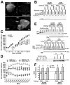
Figure 2. Overexpression of GRK6 in the motor striatum suppresses L-DOPA-induced contralateral rotations in the hemiparkinsonian rat
(A) The expression of GRK6-GFP in the rat lesioned striatum (upper image) detected with anti-GFP antibody as described in Methods. The same section was co-stained with anti-tyrosine hydroxylase (TH) antibody (lower image) to show that only the lesioned side received the GRK6 lentivirus. CPu, caudatoputamen, NAC – nucleus accumbens, Sept, septum, Ctx, cortex. (B) Loss of TH in the lesioned striatum. Upper panel shows representative Western blot for TH. Standards were produced by serial dilutions of the tissue lysate from control striata of several rats. The values are expressed in μg of total striatal protein per lane. Lower panel – blot for β-actin following stripping and re-probing the original TH blot demonstrating equal loading. (C) The graph shows net contralateral L-DOPA-induced rotations registered for 1 h in rats pre-sensitized with L-DOPA for 5 days before injection of the lentivirus encoding GFP or GRK6 and then tested with L-DOPA for 10 days post-injection. * - p<0.05, ** - p<0.01 between the GRK6 and control GFP groups, post hoc Student’s unpaired t-test following two-way repeated measure ANOVA with Protein (GFP versus GRK6) as between and Session as repeated measure factors. Note that both groups had similar rotation frequencies before virus injection, whereas after the injection GRK6-expressing rats demonstrated reduced rotational behavior. (D) The graph representing the distribution of net contralateral rotations across 5-min bins during the first (Day 1) and last (Day 10) 60 min testing sessions. * - p<0.05, ** - p<0.01, * - p<0.001 between the GRK6 and corresponding control GFP groups, post hoc Student’s unpaired t-test following two-way repeated measure ANOVA with Proteins (GFP versus GRK6) as between group and Bin as repeated measure factors. (E) The upper panel shows the expression of GFP in the lesioned striatum of GFP-expressing animals of all experimental groups. The middle panel shows the expression of endogenous GRK6 in both intact and lesioned striata of GFP-expressing rats and in the intact striatum of GRK6-overexpressing rats of all experimental groups detected with anti-GRK6 antibody. Serial dilutions of purified recombinant human GRK6 were used as standards (left 6 lanes). In the lesioned striatum of rats injected with GRK6 virus total (endogenous plus exogenous) GRK6 was detected. The arrow points to the GRK6 band (the lower thicker band is nonspecific). Lower images in both panels show blots for β-actin following stripping and re-probing the original blots to demonstrate equal loading. Lower panel shows a higher magnification of a part of the GRK6 blot to emphasize the differences in the GRK6 levels between the intact and lesioned striata in the GFP-expressing rat and between the lesioned striata in the GFP- and GRK6-expressing rats. (F) The graph presenting quantification of the levels of endogenous and total GRK6 in the lesioned caudate-putamen in rats injected with lentivirus encoding GFP or GRK6, as percent of values of respective intact hemispheres (means±S.E.M.). N=9–10 rats per group. * - p<0.01, * - p<0.05 to respective intact hemisphere by one-way repeated measure ANOVA separately for each group; # - p<0.001 to respective lesioned hemisphere in GFP-expressing rats by Student’s unpaired t-test.
Figure 3
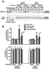
Figure 3. Overexpression of GRK6 in the striatum reduces ERK activation induced by acute L-DOPA
(A) Western blot showing activation of ERK1/2 in the lesioned striatum of rats expressing GFP (control) or GRK6, chronically treated with saline or L-DOPA and challenged with either saline or L-DOPA (upper panel). Lower blot shows total ERK1/2 expression. (B) Quantification of Western blots of ERK1/2 activation by acute L-DOPA challenge in rats expressing GFP or GRK6 in the lesioned striatum and chronically treated with either saline (drug-naïve) of L-DOPA for 10 days. Rats were challenged with either L-DOPA or saline 45 min prior to sacrifice. N=9–10 rats per group. * - p<0.001 ** - p<0.01, to intact striatum, paired Student’s t-test; a –p<0.05 – to GFP-expressing animals by two-way ANOVA separately in the lesioned striatum in L-DOPA-challenged groups across treatments; # - p<0.05 to the GFP-expressing SALINE-treated L-DOPA-challenged group, lesioned striatum, group, unpaired Student’s post hoc t-test.
Figure 4
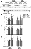
Figure 4. Overexpression of GRK6 in the striatum reduces p38 phosphorylation induced by acute L-DOPA
(A) Western blots showing activation of p38 in the lesioned striatum of rats expressing GFP (control) or GRK6, chronically treated with saline or L-DOPA, and challenged with either saline or L-DOPA (upper panel). Lower blot shows total p38 expression. (B) Western blots showing activation of p38 in the lesioned striatum of rats expressing GFP (control) or GRK6, chronically treated with saline or L-DOPA and challenged with either saline or L-DOPA. (C) Quantification of the Western blot data for total p38 expression. (D) Ratio of phospho-p38 to total p38 calculated for each animal quantified across experimental groups. N=9–10 rats per group. * - p<0.001, ** - p<0.01, * - p<0.5, to intact striatum, paired Student’s t-test; a – p<0.05 – to GFP-expressing animals by three-way ANOVA separately in the lesioned striatum across treatments and challenges; # - p<0.05 to the GFP-expressing SALINE-treated L-DOPA-challenged group, lesioned striatum, group, unpaired Student’s post hoc t-test.
Figure 5
Figure 5. Overexpression of GRK6 in the striatum has no effect on the phosphorylation or expression of JNK isoforms in hemiparkinsonian rats
Western blot images showing activation of JNK1/2 in the lesioned striatum of rats expressing GFP (control) or GRK6, chronically treated with saline or L-DOPA and challenged with either saline or L-DOPA (upper panel). Lower blot shows total JNK1/2 expression.
Figure 6
Figure 6. Overexpression of GRK6 in the lesioned striatum reduces phosphorylation of Akt at Thr308
(A) Western blots showing activation of Akt at Thr308 (upper panel) and Ser473 (middle panel) in the lesioned striatum of rats expressing GFP (control) or GRK6, chronically treated with saline or L-DOPA and challenged with either saline or L-DOPA. Lower blot shows total Akt expression. (B) Quantification of Western blots of Akt phosphorylation at Thr308 and Ser473 following acute challenge with L-DOPA or saline in rats expressing GFP or GRK6 in the lesioned striatum and treated with either saline (drug-naïve) of L-DOPA for 10 days. N=9–10 rats per group. * - p<0.001, ** - p<0.01, * - p<0.05 to intact striatum, paired Student’s t-test; a – p<0.05 – to GFP-expressing animals by two-way ANOVA separately in the lesioned striatum in L-DOPA-challenged groups across treatments.
Figure 7
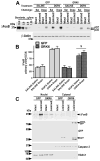
Figure 7. Overexpression of GRK6 in the lesioned striatum reduces L-DOPA-induced accumulation of ΔFosB
(A) Representative Western blots showing ΔFosB accumulation in the lesioned striatum of rats expressing GFP (control) or GRK6, chronically treated with saline or L-DOPA and challenged with either saline or L-DOPA. Note that there is no ΔFosB expression in the intact striatum regardless of treatment or challenge. Arrows point to the two ΔFosB isoforms. Lower panel – Western blot for β-actin produced by stripping the original ΔFosB blot and reprobing with β-acting antibody. (B) Quantification of Western blots of ΔFosB expression in the lesioned striatum following acute challenge with L-DOPA or saline in rats expressing GFP or GRK6 treated with either saline (drug-naïve) of L-DOPA for 10 days. N=9–10 rats per group. * - p<0.05 to GFP-expressing L-DOPA-treated groups across challenges by two-way ANOVA with Protein and Challenge. (C) Detection of ΔFosB accumulation in the nuclei as compared to the cytosolic fractions of the intact and lesioned striata of rats expressing GFP or GRK6-GFP, chronically treated with saline or L-DOPA. Nuclear and cytosolic fractions were isolated as described in Methods. Representative Western blot of nuclear and cytosolic (7.5 μg protein per lane) were probed for ΔFosB, and then blots were stripped and re-probed for caspase-3 and histone deacetylase 1 (HDAC1) to demonstrate equal loading of the samples in both fractions. Lentivirally-delivered GFP and GRK6-GFP were detected with anti-GFP antibody on a separate blot (seen only in the cytosolic fraction as expected).

Similar articles

Cited by

References

    1. Ahmed MR, Berthet A, Bychkov E, Porras G, Li Q, Bioulac BH, Carl YT, Bloch B, Kook S, Aubert I, Dovero S, Doudnikoff E, Gurevich VV, Gurevich EV, Bezard E. Lentiviral overexpression of GRK6 alleviates L-dopa-induced dyskinesia in experimental Parkinson’s disease. Sci Transl Med. 2010;2:28ra28. - PMC - PubMed
    1. Ahmed MR, Bychkov ER, Gurevich VV, Benovic JL, Gurevich EV. Altered expression and subcellular distribution of GRK subtypes in the dopamine-depleted rat basal ganglia is not normalized by L-DOPA treatment. J Neurochem. 2007;104:1622–1636. - PMC - PubMed
    1. Ahmed MR, Gurevich VV, Dalby KN, Benovic JL, Gurevich EV. Haloperidol and clozapine differentially affect the expression of arrestins, receptor kinases, and extracellular signal-regulated kinase activation. J Pharmacol Exper Ther. 2008;325:276–83. - PMC - PubMed
    1. Alcacer C, Santini E, Valjent E, Gaven F, Girault JA, Hervé D. Gα(olf) mutation allows parsing the role of cAMP-dependent and extracellular signal-regulated kinase-dependent signaling in L-3,4-dihydroxyphenylalanine-induced dyskinesia. J Neurosci. 2012;32:5900–5910. - PMC - PubMed
    1. Alessi DR, Andjelkovic M, Caudwell B, Cron P, Morrice N, Cohen P, Hemmings BA. Mechanism of activation of protein kinase B by insulin and IGF-1. EMBO J. 1996;15:6541–6551. - PMC - PubMed

Publication types

MeSH terms