

Array tomography for the detection of non-dilated, injured axons in traumatic brain injury
- PMID: 25687633
- PMCID: PMC4393800
- DOI: 10.1016/j.jneumeth.2015.02.005
Array tomography for the detection of non-dilated, injured axons in traumatic brain injury
Abstract
Background: Axonal injury is a key feature of several types of brain trauma and neurological disease. However, in mice and humans, many axons are less than 500 nm in diameter which is at or below the resolution of most conventional light microscopic imaging methods. In moderate to severe forms of axon injury, damaged axons become dilated and therefore readily detectible by light microscopy. However, in more subtle forms of injury, the damaged axons may remain undilated and therefore difficult to detect.
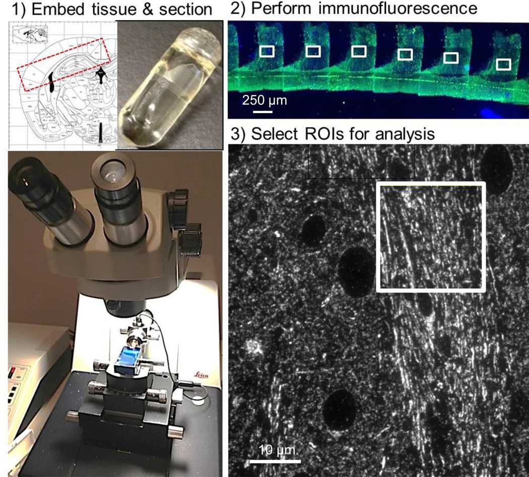
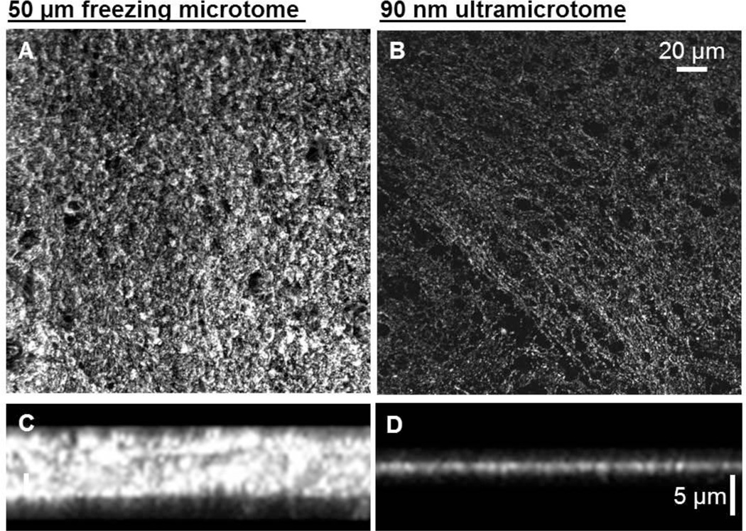
New method: Here we present a method for adapting array tomography for the identification and quantification of injured axons. In this technique, ultrathin (∼70 nm) plastic sections of tissue are prepared, labeled with axon injury-relevant antibodies and imaged using conventional epifluorescence.
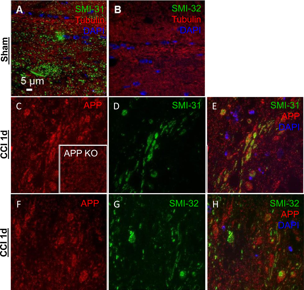
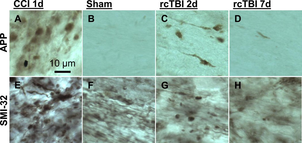
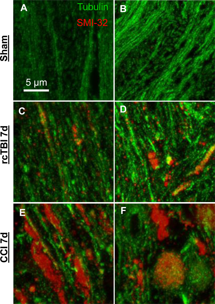
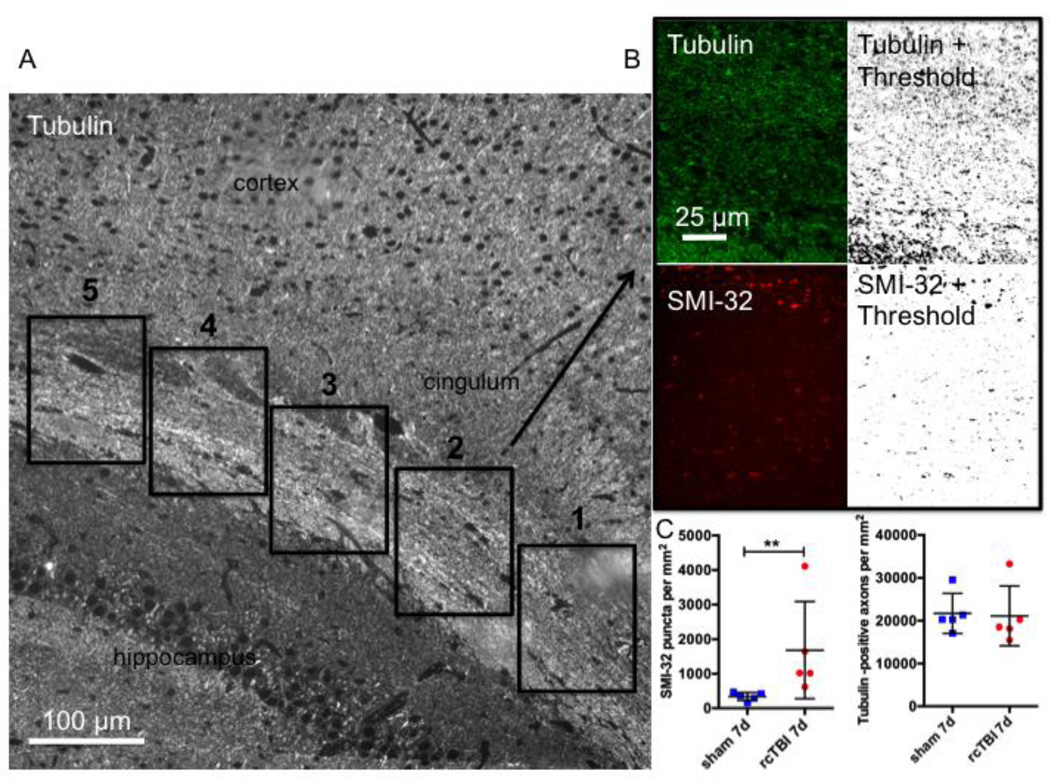
Results: To demonstrate the use of array-tomography-based methods, we determined that mice that received two closed-skull concussive traumatic brain injury impacts had significantly increased numbers of non-dilated axons that were immunoreactive for non-phosphorylated neurofilament (SMI-32; a marker of axonal injury), compared to sham mice (1682±628 versus 339±52 per mm(2), p=0.004, one-tailed Mann-Whitney U test). Tubulin loss was not evident (p=0.2063, one-tailed Mann-Whitney U test). Furthermore, mice that were subjected to more severe injury had a loss of tubulin in addition to both dilated and non-dilated SMI-32 immunoreactive axons indicating that this technique is suitable for the analysis of various injury conditions.
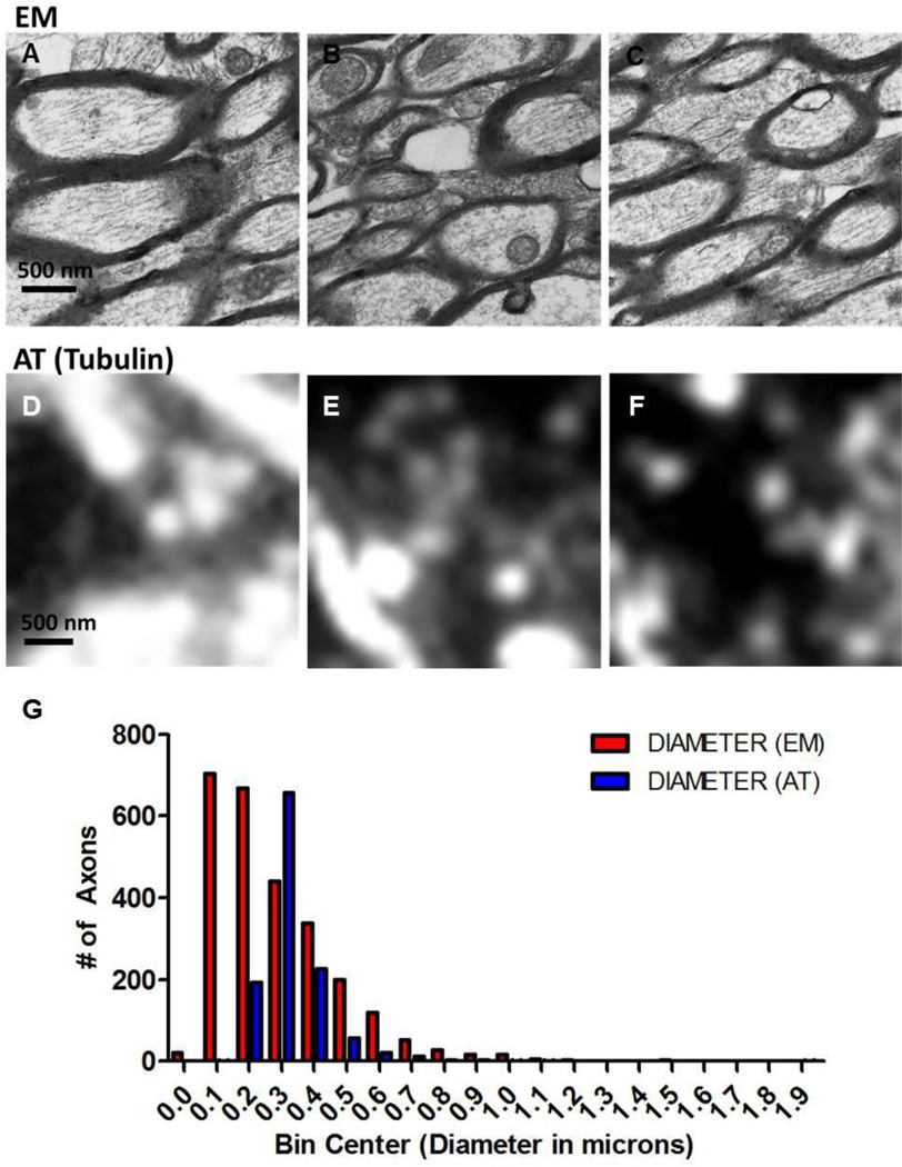
Comparison with existing method: With array tomography we could detect similar overall numbers of axons as electron microscopy, but accurate diameter measurements were limited to those with diameters >200 nm. Importantly, array tomography had greater sensitivity for detecting small non-dilated injured axons compared with conventional immunohistochemistry.
Conclusion: Imaging of individual axons and quantification of subtle axonal injury is possible using this array tomography method. This method may be most useful for the assessment of concussive injuries and other pathologies in which injured axons are not typically dilated. The ability to process moderately large volumes of tissue, label multiple proteins of interest, and automate analysis support array tomography as a useful alternative to electron microscopy.
Keywords: Array tomography; Axonal injury; Electron microscopy; Neurofilament; Traumatic brain injury; Tubulin.
Copyright © 2015 Elsevier B.V. All rights reserved.
Figures
Similar articles
-
Characterization of a prolonged regenerative attempt by diffusely injured axons following traumatic brain injury in adult cat: a light and electron microscopic immunocytochemical study.Acta Neuropathol. 1997 Oct;94(4):329-37. doi: 10.1007/s004010050715. Acta Neuropathol. 1997. PMID: 9341933
-
Mild axonal stretch injury in vitro induces a progressive series of neurofilament alterations ultimately leading to delayed axotomy.J Neurotrauma. 2005 Oct;22(10):1081-91. doi: 10.1089/neu.2005.22.1081. J Neurotrauma. 2005. PMID: 16238485
-
Repetitive closed-skull traumatic brain injury in mice causes persistent multifocal axonal injury and microglial reactivity.J Neuropathol Exp Neurol. 2011 Jul;70(7):551-67. doi: 10.1097/NEN.0b013e31821f891f. J Neuropathol Exp Neurol. 2011. PMID: 21666502 Free PMC article.
-
A mechanistic analysis of nondisruptive axonal injury: a review.J Neurotrauma. 1997 Jul;14(7):419-40. doi: 10.1089/neu.1997.14.419. J Neurotrauma. 1997. PMID: 9257661 Review.
-
Protein accumulation in traumatic brain injury.Neuromolecular Med. 2003;4(1-2):59-72. doi: 10.1385/NMM:4:1-2:59. Neuromolecular Med. 2003. PMID: 14528053 Review.
Cited by
-
Distinctive Structural and Molecular Features of Myelinated Inhibitory Axons in Human Neocortex.eNeuro. 2018 Oct 16;5(5):ENEURO.0297-18.2018. doi: 10.1523/ENEURO.0297-18.2018. eCollection 2018 Sep-Oct. eNeuro. 2018. PMID: 30406183 Free PMC article.
-
Combined [(18)F]DPA-714 micro-positron emission tomography and autoradiography imaging of microglia activation after closed head injury in mice.J Neuroinflammation. 2016 Jun 7;13(1):140. doi: 10.1186/s12974-016-0604-9. J Neuroinflammation. 2016. PMID: 27266706 Free PMC article.
-
Correcting miR92a-vGAT-Mediated GABAergic Dysfunctions Rescues Human Tau-Induced Anxiety in Mice.Mol Ther. 2017 Jan 4;25(1):140-152. doi: 10.1016/j.ymthe.2016.10.010. Epub 2017 Jan 4. Mol Ther. 2017. PMID: 28129110 Free PMC article.
-
Identifying the Phenotypes of Diffuse Axonal Injury Following Traumatic Brain Injury.Brain Sci. 2023 Nov 20;13(11):1607. doi: 10.3390/brainsci13111607. Brain Sci. 2023. PMID: 38002566 Free PMC article. Review.
-
Microglial depletion abolishes ischemic preconditioning in white matter.Glia. 2022 Apr;70(4):661-674. doi: 10.1002/glia.24132. Epub 2021 Dec 23. Glia. 2022. PMID: 34939240 Free PMC article.
References
-
- Aboitiz F, Scheibel AB, Fisher RS, Zaidel E. Fiber composition of the human corpus callosum. Brain research. 1992;598:143–153. - PubMed
-
- Blumbergs PC, Scott G, Manavis J, Wainwright H, Simpson DA, McLean AJ. Staining of amyloid precursor protein to study axonal damage in mild head injury. Lancet. 1994;344:1055–1056. - PubMed
Publication types
MeSH terms
Substances
Grants and funding
LinkOut - more resources
Full Text Sources
Other Literature Sources
