Application of IL-36 receptor antagonist weakens CCL20 expression and impairs recovery in the late phase of murine acetaminophen-induced liver injury
- PMID: 25687687
- PMCID: PMC4330543
- DOI: 10.1038/srep08521
Application of IL-36 receptor antagonist weakens CCL20 expression and impairs recovery in the late phase of murine acetaminophen-induced liver injury
Abstract
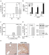
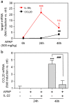
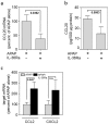
Overdosing of the analgesic acetaminophen (APAP, paracetamol) is a major cause of acute liver injury. Whereas toxicity is initiated by hepatocyte necrosis, course of disease is regulated by mechanisms of innate immunity having the potential to serve in complex manner pathogenic or pro-regenerative functions. Interleukin (IL)-36γ has been identified as novel IL-1-like cytokine produced by and targeting epithelial (-like) tissues. Herein, we investigated IL-36γ in acute liver disease focusing on murine APAP-induced hepatotoxicity. Enhanced expression of hepatic IL-36γ and its prime downstream chemokine target CCL20 was detected upon liver injury. CCL20 expression coincided with the later regeneration phase of intoxication. Primary murine hepatocytes and human Huh7 hepatocellular carcinoma cells indeed displayed enhanced IL-36γ expression when exposed to inflammatory cytokines. Administration of IL-36 receptor antagonist (IL-36Ra) decreased hepatic CCL20 in APAP-treated mice. Unexpectedly, IL-36Ra likewise increased late phase hepatic injury as detected by augmented serum alanine aminotransferase activity and histological necrosis which suggests disturbed tissue recovery upon IL-36 blockage. Finally, we demonstrate induction of IL-36γ in inflamed livers of endotoxemic mice. Observations presented introduce IL-36γ as novel parameter in acute liver injury which may contribute to the decision between unleashed tissue damage and initiation of liver regeneration during late APAP toxicity.
Figures


Similar articles
-
Application of interleukin-22 mediates protection in experimental acetaminophen-induced acute liver injury.Am J Pathol. 2013 Apr;182(4):1107-13. doi: 10.1016/j.ajpath.2012.12.010. Epub 2013 Jan 30. Am J Pathol. 2013. PMID: 23375450
-
CHOP is a critical regulator of acetaminophen-induced hepatotoxicity.J Hepatol. 2013 Sep;59(3):495-503. doi: 10.1016/j.jhep.2013.04.024. Epub 2013 May 9. J Hepatol. 2013. PMID: 23665281
-
M1 muscarinic receptors modify oxidative stress response to acetaminophen-induced acute liver injury.Free Radic Biol Med. 2015 Jan;78:66-81. doi: 10.1016/j.freeradbiomed.2014.09.032. Epub 2014 Oct 31. Free Radic Biol Med. 2015. PMID: 25452146 Free PMC article.
-
STAT3, a Key Parameter of Cytokine-Driven Tissue Protection during Sterile Inflammation - the Case of Experimental Acetaminophen (Paracetamol)-Induced Liver Damage.Front Immunol. 2016 May 2;7:163. doi: 10.3389/fimmu.2016.00163. eCollection 2016. Front Immunol. 2016. PMID: 27199988 Free PMC article. Review.
-
Protective effects of nicotinamide against acetaminophen-induced acute liver injury.Int Immunopharmacol. 2012 Dec;14(4):530-7. doi: 10.1016/j.intimp.2012.09.013. Epub 2012 Oct 9. Int Immunopharmacol. 2012. PMID: 23059795 Review.
Cited by
-
3'mRNA sequencing reveals pro-regenerative properties of c5ar1 during resolution of murine acetaminophen-induced liver injury.NPJ Regen Med. 2022 Jan 27;7(1):10. doi: 10.1038/s41536-022-00206-x. NPJ Regen Med. 2022. PMID: 35087052 Free PMC article.
-
A Prominent Role of Interleukin-18 in Acetaminophen-Induced Liver Injury Advocates Its Blockage for Therapy of Hepatic Necroinflammation.Front Immunol. 2018 Feb 8;9:161. doi: 10.3389/fimmu.2018.00161. eCollection 2018. Front Immunol. 2018. PMID: 29472923 Free PMC article.
-
Interleukin-1 Family Cytokines: Keystones in Liver Inflammatory Diseases.Front Immunol. 2019 Aug 27;10:2014. doi: 10.3389/fimmu.2019.02014. eCollection 2019. Front Immunol. 2019. PMID: 31507607 Free PMC article. Review.
-
The Protective Role of IL-36/IL-36R Signal in Con A-Induced Acute Hepatitis.J Immunol. 2022 Feb 15;208(4):861-869. doi: 10.4049/jimmunol.2100481. Epub 2022 Jan 19. J Immunol. 2022. PMID: 35046104 Free PMC article.
-
Selective targeting of tumor associated macrophages in different tumor models.PLoS One. 2018 Feb 15;13(2):e0193015. doi: 10.1371/journal.pone.0193015. eCollection 2018. PLoS One. 2018. PMID: 29447241 Free PMC article.
References
-
- Bernau W., Auzinger G., Dhawan A. & Wendon J. Acute liver failure. Lancet 376, 190–201 (2010). - PubMed
-
- Kubes P. & Mehal W. Z. Sterile inflammation in the liver. Gastroenterology 143, 1158–1172 (2012). - PubMed
-
- Brenner C., Galluzzi L., Kepp O. & Kroemer G. Decoding cell death signals in liver inflammation. J. Hepatol. 59, 583–594 (2013). - PubMed
Publication types
MeSH terms
Substances
LinkOut - more resources
Full Text Sources
Other Literature Sources
Medical

