. 2015 Feb 17:6:6281.
doi: 10.1038/ncomms7281.

The polymeric mucin Muc5ac is required for allergic airway hyperreactivity

Affiliations

The polymeric mucin Muc5ac is required for allergic airway hyperreactivity

Christopher M Evans et al. Nat Commun. .

Abstract

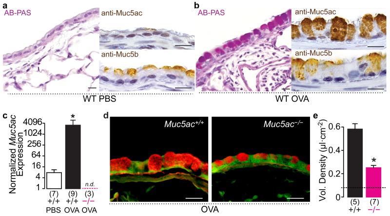
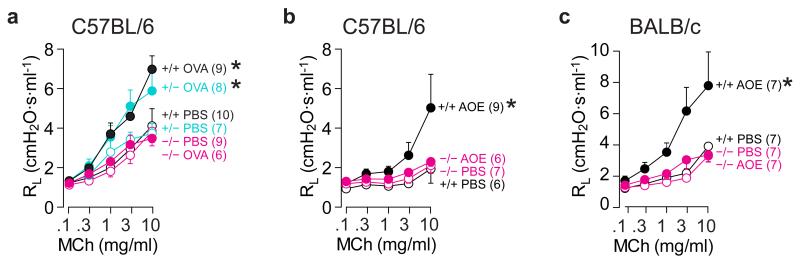
In asthma, airflow obstruction is thought to result primarily from inflammation-triggered airway smooth muscle (ASM) contraction. However, anti-inflammatory and smooth muscle-relaxing treatments are often temporary or ineffective. Overproduction of the mucin MUC5AC is an additional disease feature that, while strongly associated pathologically, is poorly understood functionally. Here we show that Muc5ac is a central effector of allergic inflammation that is required for airway hyperreactivity (AHR) to methacholine (MCh). In mice bred on two well-characterized strain backgrounds (C57BL/6 and BALB/c) and exposed to two separate allergic stimuli (ovalbumin and Aspergillus extract), genetic removal of Muc5ac abolishes AHR. Residual MCh responses are identical to unchallenged controls, and although inflammation remains intact, heterogeneous mucous occlusion decreases by 74%. Thus, whereas inflammatory effects on ASM alone are insufficient for AHR, Muc5ac-mediated plugging is an essential mechanism. Inhibiting MUC5AC may be effective for treating asthma and other lung diseases where it is also overproduced.


Figures

Fig. 1
Fig. 1. Mucous metaplasia is significantly reduced in Muc5ac−/− airways
(a and b) Mucin production in saline (a) and OVA (b) challenged WT mice. AB-PAS, anti-Muc5ac, and anti-MUC5B staining shows up-regulated Muc5ac and sustained MUC5B in allergic airways. (c-e) Muc5ac mRNA increased 383-fold in OVA challenged WT mice (+/+), but it was undetectable in Muc5ac knockout (−/−) mice (c), resulting in a 67% reduction in mucous metaplasia detected by periodic acid fluorescent Schiff’s staining (d and e). Scale bars, 20 μm (a, b, and d). Mean, s.e.m. (c and e). ‘*’, p<0.05 (one-tailed) by unpaired t-test (c and e), with Welch’s correction for unequal variances in c. ‘n.d.’, not detected. Numbers in parentheses, ‘N’ mice. ‘Vol.’, volume. Dashed line in e, baseline in saline challenged WT mice.
Fig. 2
Fig. 2. Muc5ac is required for AHR to MCh
Lung resistance (RL) responses to MCh in WT (+/+, black), Muc5ac+/- (turquoise), and Muc5ac−/− (magenta) mice after saline (open circles), OVA (closed circles in a), or Aspergillus oryzae extract (AOE; closed circles in b and c). Mean, s.e.m. (a-c). ‘*’, p<0.05 between slopes of best-fit regression lines by one-way ANOVA. Numbers in parentheses, ‘N’ mice.
Fig. 3
Fig. 3. Muc5ac mediates mouse strain-specific allergic airway pathophysiology
Conducting airway resistance (ΔRAW), tissue resistance (ΔGTI) and tissue elastance (ΔHTI) were measured using oscillatory mechanics to assess input impedance. (a) In WT C57BL/6 mice, Aspergillus oryzae extract (AOE)-induced AHR (closed black circles) was dominated by changes in peripheral tissue mechanics (ΔGTI and ΔHTI) compared to changes in RAW. (b) In WT BALB/c mice, AOE-induced AHR (closed black circles) had central airway (ΔRAW) and tissue dominance (ΔGTI), whereas ΔHTI was not significantly different following AOE (n.s.) in WT mice. In both congenic strains, responses were significantly blunted in Muc5ac−/− mice (closed magenta circles). Mean, s.e.m. ‘*’, p<0.05 between slopes of best-fit regression lines by one-way ANOVA. ‘n.s.’, not significant. Numbers in parentheses, ‘N’ mice.
Fig. 4
Fig. 4. Acute mucin secretion does not cause AHR
WT mice were OVA or PBS challenged, and then anesthetized, ventilated, and exposed to UTP (0.1-100 mg/ml) or vehicle (saline). (a) In OVA challenged lungs fixed immediately after dose response tests, there was an acute decrease in periodic acid fluorescent Schiff’s (PAFS) staining in UTP (right panel) compared to vehicle (‘-UTP’, left panel) treated mice, demonstrating acute UTP-induced secretion. Scale bars, 20 μm. (b) Quantitation of UTP-induced changes in PAFS staining in OVA challenged mice. (c) No effect of UTP on RL in either PBS or OVA challenged WT mice. Mean, s.e.m. in b and c. Numbers in parentheses, ‘N’ mice. ‘Vol.’, volume. Dashed line in b, baseline in saline challenged WT mice. ‘*’, p < 0.05 by t Test.
Fig. 5
Fig. 5. Muc5ac deficiency does not reduce allergic inflammation
WT (black) and Muc5ac−/− (red) mice had similar numbers of eosinophils in lung lavage fluid irrespective of OVA or Aspergillus oryzae extract (AOE) antigen challenge or strain background variables. For all groups, saline challenged mice had <104 eosinophils in lung lavage fluid (see Supplementary Table 1). Mean, s.e.m. N = 6-11 mice/group.
Fig. 6
Fig. 6. Muc5ac deficiency protects against heterogeneous airflow obstruction
(a) Models of the effects of uniform vs. non-uniform obstruction on airflow. (b and c) Changes in peak total lung and airway resistances (ΔR) following 10 mg/ml MCh in WT and Muc5ac−/− mice (n = 7 per genotype) (b) were fit to Poiseuille’s Law (equation in c) to predict changes in airway lumen diameter (red point and dashed arrow). Viscosity (η) and length (l) were held constant. (d-f) Mean (d) and distributions (e,f) of airway lumen diameters measured in methacarn fixed lungs; changes in mean lumen diameter were also fit to the equation in c (black points and dashed arrow). (g-i) Contributions of mucus, airway epithelium, and smooth muscle to changes in composite airway wall/mucus (AW/M) thickness. Mean, s.e.m. in b, d, g-i. x-axis labels bin maximum values in e, f. N = 4 WT and 5 Muc5ac−/− mice. ‘*’, p < 0.05 by t Test.
Fig. 7
Fig. 7. Muc5ac deficiency reduces mucus plugging
(a and b) Inhaled MCh caused mucus plugging in WT allergic mouse airways (aw). Muc5ac protein (a) and PAFS staining (b) in AOE challenged WT (Muc5ac+/+) and Muc5ac−/− mouse airways. Arrow in A, region shown at higher magnification in right panel. (c-f) Extent and distribution of mucus plugging. Mean occlusion per mouse across all measured airways (c). Distribution of the extent (d) and localization (e and f) of occlusions. Arrows in e and f, starts of upper quartiles. Scale bars, 100 and 20 μm (a); 50 μm (b). Mean, s.e.m. (c). Median-quartiles (d). ‘*’, p<0.05 (one-tailed) by unpaired t-test (c) and Mann-Whitney test (d). Numbers in parentheses, ‘N’ mice (c), ‘N’ airways (d-f).

References

    1. Fanta CH. Asthma. N Engl J Med. 2009;360:1002–1014. doi:10.1056/NEJMra0804579. - PubMed
    1. Bosse Y, Riesenfeld EP, Pare PD, Irvin CG. Annu Rev Physiol. 2010;72:437–462. doi:10.1146/annurev-physiol-021909-135851. - PubMed
    1. Wagers SS, et al. Extravascular fibrin, plasminogen activator, plasminogen activator inhibitors, and airway hyperresponsiveness. J Clin.Invest. 2004;114:104–111. - PMC - PubMed
    1. Fahy JV, Dickey BF. Airway mucus function and dysfunction. New England Journal of Medicine. 2010;363:2233–2247. - PMC - PubMed
    1. Lundblad LK, et al. Airway hyperresponsiveness in allergically inflamed mice: the role of airway closure. Am J Respir Crit Care Med. 2007;175:768–774. - PMC - PubMed

Publication types