

Cerebrospinal fluid leakage and headache after lumbar puncture: a prospective non-invasive imaging study
- PMID: 25688077
- PMCID: PMC4614121
- DOI: 10.1093/brain/awv016
Cerebrospinal fluid leakage and headache after lumbar puncture: a prospective non-invasive imaging study
Abstract
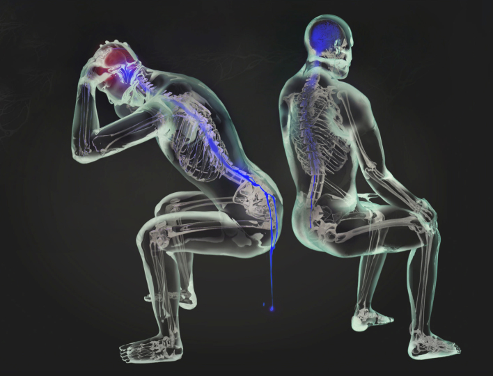
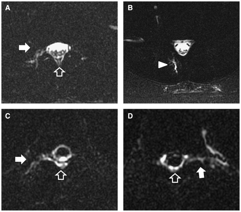
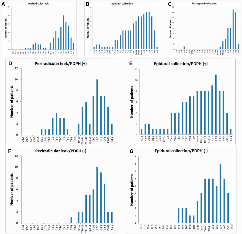
The spatial distribution and clinical correlation of cerebrospinal fluid leakage after lumbar puncture have not been determined. Adult in-patients receiving diagnostic lumbar punctures were recruited prospectively. Whole-spine heavily T2-weighted magnetic resonance myelography was carried out to characterize post-lumbar puncture spinal cerebrospinal fluid leakages. Maximum rostral migration was defined as the distance between the most rostral spinal segment with cerebrospinal fluid leakage and the level of lumbar puncture. Eighty patients (51 female/29 male, mean age 49.4 ± 13.3 years) completed the study, including 23 (28.8%) with post-dural puncture headache. Overall, 63.6% of periradicular leaks and 46.9% of epidural collections were within three vertebral segments of the level of lumbar puncture (T12-S1). Post-dural puncture headache was associated with more extensive and more rostral distributions of periradicular leaks (length 3.0 ± 2.5 versus 0.9 ± 1.9 segments, P = 0.001; maximum rostral migration 4.3 ± 4.7 versus 0.8 ± 1.7 segments, P = 0.002) and epidural collections (length 5.3 ± 6.1 versus 1.0 ± 2.1 segments, P = 0.003; maximum rostral migration 4.7 ± 6.7 versus 0.9 ± 2.4 segments, P = 0.015). In conclusion, post-dural puncture headache was associated with more extensive and more rostral distributions of periradicular leaks and epidural collections. Further, visualization of periradicular leaks was not restricted to the level of dural defect, although two-thirds remained within the neighbouring segments.
Keywords: cerebrospinal fluid leakage; magnetic resonance myelography; post-dural puncture headache; spontaneous intracranial hypotension.
© The Author (2015). Published by Oxford University Press on behalf of the Guarantors of Brain. All rights reserved. For Permissions, please email: journals.permissions@oup.com.
Figures
Similar articles
-
Incidental findings of CSF leakage in patients without spontaneous intracranial hypotension and development of post-dural puncture headache.Eur Radiol. 2014 Apr;24(4):827-33. doi: 10.1007/s00330-013-3070-0. Epub 2013 Nov 22. Eur Radiol. 2014. PMID: 24272226
-
Ventral spinal cerebrospinal fluid leak as the cause of persistent post-dural puncture headache in children.J Neurosurg Pediatr. 2013 Jan;11(1):48-51. doi: 10.3171/2012.10.PEDS12353. Epub 2012 Nov 9. J Neurosurg Pediatr. 2013. PMID: 23140214
-
Pediatric post-dural puncture headache and paraplegia.Headache. 2024 Jul-Aug;64(7):865-868. doi: 10.1111/head.14749. Epub 2024 Jun 11. Headache. 2024. PMID: 38860510
-
[Post-dural puncture headache and blood-patch: theoretical and practical approach].Ann Fr Anesth Reanim. 2013 May;32(5):325-38. doi: 10.1016/j.annfar.2013.02.014. Epub 2013 Apr 6. Ann Fr Anesth Reanim. 2013. PMID: 23566592 Review. French.
-
[Post-lumbar puncture syndrome--its pathogenesis, prophylaxis and treatment].Neurol Neurochir Pol. 2006 Sep-Oct;40(5):434-40. Neurol Neurochir Pol. 2006. PMID: 17103357 Review. Polish.
Cited by
-
The ebb and flow of headache: A clue to pathophysiology of sinus stenosis in idiopathic intracranial hypertension?J Postgrad Med. 2023 Jul-Sep;69(3):179-181. doi: 10.4103/jpgm.jpgm_238_22. J Postgrad Med. 2023. PMID: 36453388 Free PMC article. No abstract available.
-
Investigation of the optimal duration of bed rest in the supine position to reduce complications after lumbar puncture combined with intrathecal chemotherapy: a multicenter prospective randomized controlled trial.Support Care Cancer. 2018 Sep;26(9):2995-3002. doi: 10.1007/s00520-018-4142-0. Epub 2018 Mar 15. Support Care Cancer. 2018. PMID: 29546527 Free PMC article. Clinical Trial.
-
Comparing 2-dimensional versus 3-dimensional MR myelography for cerebrospinal fluid leak detection.Eur J Radiol Open. 2024 Apr 25;12:100565. doi: 10.1016/j.ejro.2024.100565. eCollection 2024 Jun. Eur J Radiol Open. 2024. PMID: 38699593 Free PMC article.
-
Magnetic resonance imaging predicted the therapeutic response of patients with spinal cerebrospinal fluid leakage undergoing targeted epidural blood patch.Br J Radiol. 2022 Jan 1;95(1129):20210841. doi: 10.1259/bjr.20210841. Epub 2021 Nov 22. Br J Radiol. 2022. PMID: 34762485 Free PMC article.
-
Deep learning-based reconstruction for 3-dimensional heavily T2-weighted fat-saturated magnetic resonance (MR) myelography in epidural fluid detection: image quality and diagnostic performance.Quant Imaging Med Surg. 2024 Sep 1;14(9):6531-6542. doi: 10.21037/qims-24-455. Epub 2024 Aug 7. Quant Imaging Med Surg. 2024. PMID: 39281122 Free PMC article.
References
-
- Beards SC, Jackson A, Griffiths AG, Horsman EL. Magnetic resonance imaging of extradural blood patches: appearances from 30 min to 18 h. Br J Anaesth. 1993;71:182–8. - PubMed
-
- Bezov D, Lipton RB, Ashina S. Post-dural puncture headache: part I diagnosis, epidemiology, etiology, and pathophysiology. Headache. 2010a;50:1144–52. - PubMed
-
- Bezov D, Ashina S, Lipton R. Post-dural puncture headache: Part II–prevention, management, and prognosis. Headache. 2010b;50:1482–98. - PubMed
Publication types
MeSH terms
LinkOut - more resources
Full Text Sources
Other Literature Sources
