Randomized Controlled Trial
. 2015 Apr;138(Pt 4):1009-22.
doi: 10.1093/brain/awv006. Epub 2015 Feb 15.

Rapidly progressive Alzheimer's disease features distinct structures of amyloid-β

Affiliations
Randomized Controlled Trial

Rapidly progressive Alzheimer's disease features distinct structures of amyloid-β

Mark L Cohen et al. Brain. 2015 Apr.

Abstract

Genetic and environmental factors that increase the risk of late-onset Alzheimer disease are now well recognized but the cause of variable progression rates and phenotypes of sporadic Alzheimer's disease is largely unknown. We aimed to investigate the relationship between diverse structural assemblies of amyloid-β and rates of clinical decline in Alzheimer's disease. Using novel biophysical methods, we analysed levels, particle size, and conformational characteristics of amyloid-β in the posterior cingulate cortex, hippocampus and cerebellum of 48 cases of Alzheimer's disease with distinctly different disease durations, and correlated the data with APOE gene polymorphism. In both hippocampus and posterior cingulate cortex we identified an extensive array of distinct amyloid-β42 particles that differ in size, display of N-terminal and C-terminal domains, and conformational stability. In contrast, amyloid-β40 present at low levels did not form a major particle with discernible size, and both N-terminal and C- terminal domains were largely exposed. Rapidly progressive Alzheimer's disease that is associated with a low frequency of APOE e4 allele demonstrates considerably expanded conformational heterogeneity of amyloid-β42, with higher levels of distinctly structured amyloid-β42 particles composed of 30-100 monomers, and fewer particles composed of < 30 monomers. The link between rapid clinical decline and levels of amyloid-β42 with distinct structural characteristics suggests that different conformers may play an important role in the pathogenesis of distinct Alzheimer's disease phenotypes. These findings indicate that Alzheimer's disease exhibits a wide spectrum of amyloid-β42 structural states and imply the existence of prion-like conformational strains.

Keywords: Alzheimer; progression rate; structure; β-amyloid.


Figures

Figure 1
Figure 1
Rapid rates of progression in Alzheimer’s disease are linked to low frequency of e4 allele in APOE gene but the end-point extent of amyloid or neurofibrillary tangles deposits is similar. (A) Kaplan-Meier cumulative survival analysis of cases with pathologically verified Alzheimer’s disease (AD) that were initially referred to National Prion Disease Pathology Surveillance Centre (NPDPSC) with rapidly progressive dementia (n = 186) and cases of Alzheimer’s disease (n = 2605) collected at National Alzheimer’s Coordinating Centre (NACC) database at University of Washington (Beekly et al., 2007). Statistical significance for difference in survival at ***P < 0.001 was determined with the log rank (Mantel-Cox) and generalized Wilcoxon test. (B) Frequency of e4 allele of APOE gene allelic polymorphisms in rapidly (n = 26) and slowly (n = 18) progressive cases of Alzheimer’s disease. Statistical significance at *P < 0.05 was determined with two-tailed Fisher’s exact test. (C) Typical sections of precuneus/posterior cingulate cortex (PPC), hippocampus (Hip), and cerebellum (Cer) from patients with rapidly (left) and slowly progressive Alzheimer’s disease (right). Immunohistochemistry was performed with anti-amyloid-β monoclonal antibody (BAM-10). Granule neurons of the dentate gyrus (Hip) and cerebellar granular cell layer (Cer) are designated by asterisk. Internal scale bars = 100 µm. (D) Severity of pathology classified according National Institute on Aging – Alzheimer’s Association guidelines for the neuropathologic assessment (Montine et al., 2012) in rapidly (n = 24) and slowly (n = 18) progressive cases of Alzheimer’s disease. rpAD = rapidly progressing Alzheimer’s disease; spAD = slowly progressing Alzheimer’s disease.
Figure 2
Figure 2
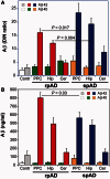
The cases with rapidly progressive Alzheimer’s disease accumulate amyloid-β42 structures with more exposed N- and C-terminal domains in cingulate and hippocampal cortex. (A) The D/N ratio was calculated from the AlphaLISA signal before (native, N) and after denaturation (denatured, D) of amyloid-β42 and amyloid-β40 in precuneus/posterior cingulate cortex (PPC), hippocampus (Hip), and cerebellum (Cer) of rapidly progressive Alzheimer’s disease (n = 26) and slowly progressive Alzheimer’s disease (n = 18) cases. The cases in which neuropathologic assessment ruled out Alzheimer’s disease, prion, or other neurodegenerative diseases were used as age-matched non-Alzheimer’s disease controls (Contr) (n = 8). The denatured state is a reference and the lower ratio indicates more exposed N- and C-terminal epitopes in native structures due to the smaller particles, different conformation, or both (Safar et al., 1998; Kim et al., 2011, 2012). (B) The levels of amyloid-β42 and amyloid-β40 in 10% homogenate of the parietal posterior cingulate cortex (PPC), hippocampus (Hip), and cerebellum (Cer) were obtained from samples denatured with 7 M Gdn HCl at 80°C. Each sample was measured in triplicate and the concentration was determined by AlphaLISA-formatted CDI calibrated with synthetic amyloid-β peptides. The bars are mean ± SEM for each parameter and statistical significance was determined with ANOVA. rpAD = rapidly progressing Alzheimer’s disease; spAD = slowly progressing Alzheimer’s disease.
Figure 3
Figure 3
Structural heterogeneity and conformationally distinct subpopulations of amyloid-β42. Structural heterogeneity and conformationally distinct subpopulations of amyloid-β42 in hippocampus of Alzheimer’s disease determined directly in the brain tissue homogenate with conformational stability assay (Safar et al., 1998; Kim et al., 2011, 2012; Safar, 2012a, b). (A) Conformational stability curves of hippocampal amyloid-β42 in rapidly progressive Alzheimer’s disease (n = 10), each curve representing dissociation and unfolding of amyloid-β42 in individual patients; (B) conformational stability curves of hippocampal amyloid-β42 of individual cases with slowly progressive Alzheimer’s disease (n = 10); (C) differential stability curves of hippocampal amyloid-β42 in rapidly progressive and slowly progressive Alzheimer’s disease fitted with Gaussian model. (D) The higher levels of unstable hippocampal amyloid-β42 conformers that are unfolding at 5.5 M Gdn HCl correlate with rapid progression of Alzheimer’s disease. The values of apparent fractional change (Fapp) of each brain sample from native to denatured state are mean ± SEM obtained from triplicate measurements at each concentration of denaturant (Gdn HCl). The analysis was performed with non-linear regression and the statistical significance was determined with ANOVA. rpAD = rapidly progressing Alzheimer’s disease; spAD = slowly progressing Alzheimer’s disease.
Figure 4
Figure 4
Particles of different sizes composed of amyloid-β42 but not amyloid-β40 in the hippocampal cortex of rapidly progressive Alzheimer’s disease (n = 10), slowly progressive Alzheimer’s disease (n = 10), and age-matched non-Alzheimer’s disease controls (n = 4). The samples were fractionated by sedimentation velocity using ultracentrifugation in sucrose gradient and analysed by CDI-formatted AlphaLISA. (A, D and G) Schematic representation of sedimentation velocity profile in sucrose gradient calibrated with standard proteins and synthetic amyloid-β42 in monomeric, oligomeric, and fibrillar state (Supplementary Fig. 3). (B, E and H) Exposure of N- and C-terminal domains in distinct particle assemblies of amyloid-β42 and amyloid-β40 monitored with D/N ratio in rapidly progressive Alzheimer’s disease (n = 10), slowly progressive Alzheimer’s disease (n = 10), and age-matched controls (n = 4). (C, F and I) Relative distribution of particles of amyloid-β42 and amyloid-β40 according to sedimentation velocity in calibrated sucrose gradient. The CDI was performed in duplicate or triplicate for each sucrose fraction of each Alzheimer’s disease case sample and the points and bars are average ± SEM. Statistical significance at *P < 0.05 and **P < 0.01 was determined with ANOVA. rpAD = rapidly progressing Alzheimer’s disease; spAD = slowly progressing Alzheimer’s disease.
Figure 5
Figure 5
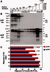
Preponderance of amyloid-β oligomeric species in rapidly progressive Alzheimer’s disease. The comparative western blots of sucrose gradient fractions from hippocampus and precuneus/posterior cingulate cortex in rapidly progressive Alzheimer’s disease (n = 12) and slowly progressive Alzheimer’s disease (n = 12). (A and B) Calibration and typical western blot of sucrose gradient fractions of rapidly progressive and slowly progressive Alzheimer’s disease cases with biotinylated mAb 6E10. The asterisk indicates floating APP100; arrows (<) point to the bands of a proteins cross-reacting with streptavidin-peroxidase complex. (C) The relative proportion of major bands of amyloid-β oligomers in top three floating fractions (#10-8) were compared with total density of a given band in all fractions from hippocampus and precuneus/posterior cingulate cortex in western blots of rapidly progressive Alzheimer’s disease (n = 12) and slowly progressive Alzheimer’s disease (n = 12). The densitometry was performed with ImageJ software and the bars represent cumulative average ± SEM for each band; the molecular mass of the markers is in kDa. Statistical significance at *P < 0.05, **P < 0.01, and ***P < 0.001 was determined with ANOVA. rpAD = rapidly progressing Alzheimer’s disease; spAD = slowly progressing Alzheimer’s disease.

Similar articles

Cited by

References

    1. Akiyama H, Mori H, Saido T, Kondo H, Ikeda K, McGeer PL. Occurrence of the diffuse amyloid beta-protein (Abeta) deposits with numerous Abeta-containing glial cells in the cerebral cortex of patients with Alzheimer’s disease. Glia. 1999;25:324–31. - PubMed
    1. Amieva H, Mokri H, Le Goff M, Meillon C, Jacqmin-Gadda H, Foubert-Samier A, et al. Compensatory mechanisms in higher-educated subjects with Alzheimer’s disease: a study of 20 years of cognitive decline. Brain. 2014;137(Pt 4):1167–75. - PubMed
    1. Beekly DL, Ramos EM, Lee WW, Deitrich WD, Jacka ME, Wu J, et al. The National Alzheimer’s Coordinating Center (NACC) database: the Uniform Data Set. Alzheimer Dis Assoc Disord. 2007;21:249–58. - PubMed
    1. Bellon A, Seyfert-Brandt W, Lang W, Baron H, Groner A, Vey M. Improved conformation-dependent immunoassay: suitability for human prion detection with enhanced sensitivity. J Gen Virol. 2003;84:1921–5. - PubMed
    1. Benilova I, Karran E, De Strooper B. The toxic Abeta oligomer and Alzheimer’s disease: an emperor in need of clothes. Nat Neurosci. 2012;15:349–57. - PubMed

Publication types