. 2015 Apr 3;290(14):9122-34.
doi: 10.1074/jbc.M114.591545. Epub 2015 Feb 16.

Phosphorylation of targeting protein for Xenopus kinesin-like protein 2 (TPX2) at threonine 72 in spindle assembly

Affiliations

Phosphorylation of targeting protein for Xenopus kinesin-like protein 2 (TPX2) at threonine 72 in spindle assembly

Su Yeon Shim et al. J Biol Chem. .

Abstract

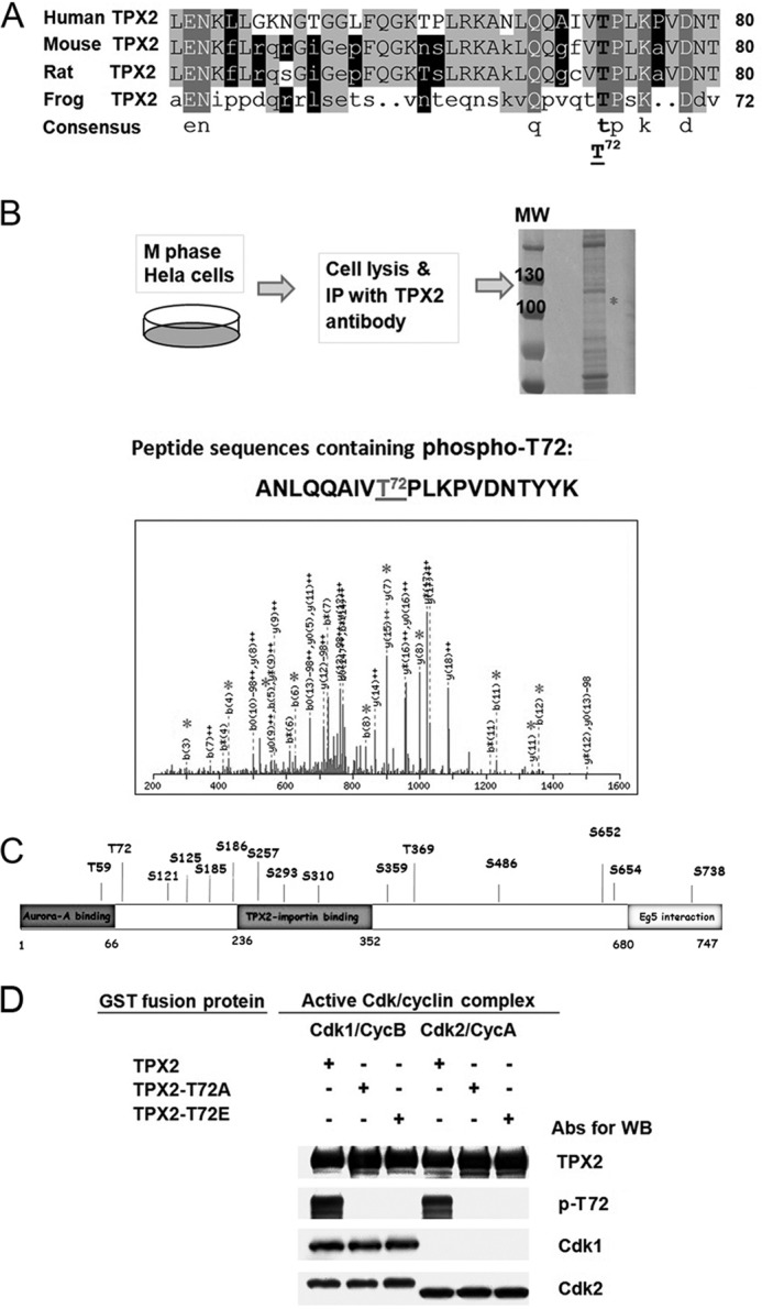
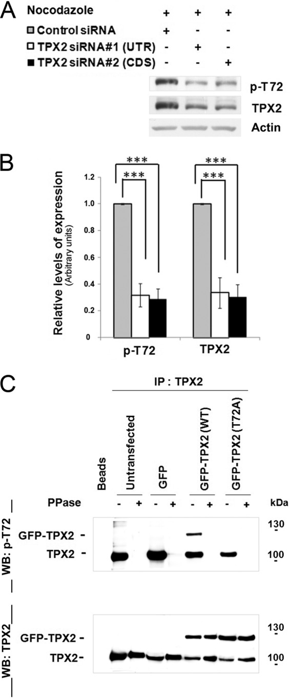
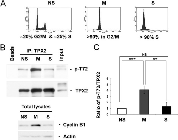
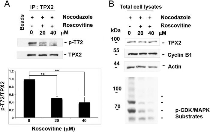
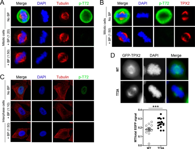
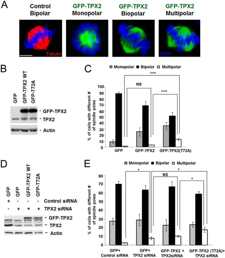
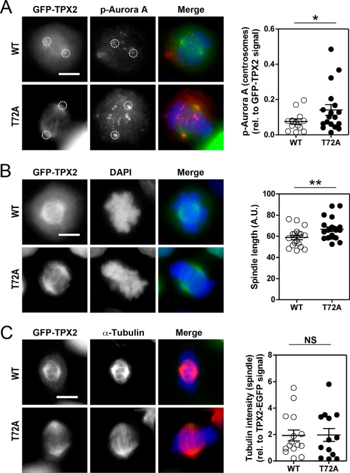
The human ortholog of the targeting protein for Xenopus kinesin-like protein 2 (TPX2) is a cytoskeletal protein that plays a major role in spindle assembly and is required for mitosis. During spindle morphogenesis, TPX2 cooperates with Aurora A kinase and Eg5 kinesin to regulate microtubule organization. TPX2 displays over 40 putative phosphorylation sites identified from various high-throughput proteomic screenings. In this study, we characterize the phosphorylation of threonine 72 (Thr(72)) in human TPX2, a residue highly conserved across species. We find that Cdk1/2 phosphorylate TPX2 in vitro and in vivo. Using homemade antibodies specific for TPX2 phosphorylated at Thr(72), we show that this phosphorylation is cell cycle-dependent and peaks at M phase. Endogenous TPX2 phosphorylated at Thr(72) does not associate with the mitotic spindle. Furthermore, ectopic GFP-TPX2 T72A preferentially concentrates on the spindle, whereas GFP-TPX2 WT distributes to both spindle and cytosol. The T72A mutant also increases the proportion of cells with multipolar spindles phenotype. This effect is associated with increased Aurora A activity and abnormally elongated spindles, indicative of higher Eg5 activity. In summary, we propose that phosphorylation of Thr(72) regulates TPX2 localization and impacts spindle assembly via Aurora A and Eg5.

Keywords: Antibody; Cell Cycle; Microtubule-associated Protein (MAP); Mitotic Spindle; Protein Phosphorylation.


Figures

FIGURE 1.
FIGURE 1.
The evolutionary conserved Thr72 in human TPX2 is phosphorylated by Cdk1 and Cdk2 in vitro. A, comparative alignment of part of human TPX2 sequence (amino acids 41 to 80) with corresponding sequences from other species (mouse, rat, and frog) using DNAMAN software (Lynnon Corporation). The sequence alignment shows that Thr72 in human TPX2 is conserved in all other species. B, schematic diagram of the experimental protocol for mass spectrometry analysis (LC-MS/MS) using mitotic HeLa cells. HeLa cells were synchronized at M phase by nocodazole treatment (100 ng/ml) for 16 h and released for 30 min after nocodazole washout. Endogenous TPX2 was immunoprecipitated from 10 mg of total protein using pan-TPX2 Abs (clone 184). IP sample was run on SDS-PAGE and after Coomassie Blue staining, the band with the matching size to TPX2 (confirmed by Western blotting with TPX2 Abs, not shown) was cut out and sent for LC-MS/MS analysis. The gray asterisk on the spectra of the phosphopeptide containing Thr72 indicate the identified matched fragment ions on mass spectrometry. C, phosphorylation sites identified by mass spectrometry analysis on endogenous TPX2 immunoprecipitated from nocodazole-synchronized mitotic HeLa cells in regards to the known TPX2 domains. All these sites have been identified previously (17, 19–32) but not confirmed and analyzed. Thr72 is the first validated and functionally characterized phosphorylation site in human TPX2 (this study). D, in vitro kinase assay using purified Cdk1/2 proteins and phosphospecific Thr72 TPX2 Abs. Purified GST fusion protein TPX2 WT, GST-TPX2-T72A, or GST-TPX2-T72E was incubated with each active Cdk-cyclin complex in the presence of 1 mm cold ATP. All kinase reactions were stopped by adding 2× SDS sample buffer and the samples were run on SDS-polyacrylamide gel electrophoresis followed by Western blot detection using the indicated antibodies.
FIGURE 2.
FIGURE 2.
Thr(P)72 TPX2 antibodies are specific in Western blot for TPX2 phosphorylated at Thr72in vivo. A, specificity of Thr(P)72 TPX2 for TPX2 protein tested by siRNA. HeLa cells were transfected with control siRNA or one of two TPX2 siRNAs for 24 h and synchronized at M phase with nocodazole treatment (100 ng/ml). Cells were harvested and lysed with lysis buffer. Samples were run on SDS-PAGE, followed by Western blotting, first probed with the Thr(P)72 TPX2 Abs, then stripped and re-probed with pan-TPX2 (clone 184) Abs. Levels of actin were used as loading controls. B, bar graph quantitation for the relative expression levels of Thr(P)72 TPX2 and TPX2 in control and TPX2 siRNA-transfected cells. Each sample was compared with sample treated with control siRNA. Relative expression levels of Thr(P)72 for control siRNA, 1 ± 0; TPX2 siRNA #1 (UTR), 0.318 ± 0.085; TPX2 siRNA #2 (Cds), 0.289 ± 0.115. Relative expression levels of TPX2, control siRNA, 1 ± 0; TPX2 siRNA #1, 0.335 ± 0.0074; TPX2 siRNA #2, 0.304 ± 0.091 (mean ± S.E.). n = 4 samples, from 4 independent experiments. Unpaired Student's t test indicated all the results are significant. ***, p < 0.001. C, specificity of Thr(P)72 TPX2 tested by the use of T72A mutant and λ-PPase treatment. HeLa cells were left untransfected, transfected with an empty GFP vector, GFP-TPX2 WT, or GFP-TPX2 T72A mutant plasmids. 24 h after transfection, cells were synchronized with nocodazole for 16 h, harvested, and lysed. TPX2 immunoprecipitation was performed in each sample with TPX2 Abs (clone 183). Where indicated, IP beads were treated with λ-PPase before SDS-PAGE. The blot was first probed with the Thr(P)72 TPX2 Abs. After stripping, the same blot was re-probed with pan-TPX2 Abs (clone 184).
FIGURE 3.
FIGURE 3.
In vivo TPX2 phosphorylation at Thr72 is cell cycle-dependent and peaks at M phase. A, cell cycle profiles analyzed by flow cytometry analysis to confirm cell synchronization at each phase. B, after IPs with pan-TPX2 Abs (clone 184), Western blots were probed first with the Thr(P)72 TPX2 Abs and then, after stripping, re-probed with pan-TPX2 Abs (clone 184). The levels of α-actin were used as loading controls. The levels of cyclin B1 were used as positive controls to show that synchronization at M phase worked well, as indicated by the high level of cyclin B1 in M phase compared with that of S phase or non-synchronized cells. The Western blot figures are representative of 3 independent experiments. The input blot is from the mitotic samples. C, bar graphs for quantification of relative levels of Thr72 phosphorylation are shown. Non-syn, non-synchronized cells (1 ± 0); M, M phase cells (4.16 ± 0.36); S, S phase cells (1.31 ± 0.4); mean of the relative levels of Thr(P)72 TPX2/TPX2 expression ± S.D., n = 3 independent experiments; ***, non-syn versus M phase, p < 0.001; **, M versus S phase, p < 0.01; NS, not significant: non-syn versus S phase, all by unpaired Student's t test.
FIGURE 4.
FIGURE 4.
Phosphorylation of TPX2 at Thr72 is inhibited by the Cdk inhibitor roscovitine. A, the levels of Thr(P)72 were reduced by roscovitine treatment. HeLa cells were first treated with 100 ng/ml of nocodazole for 16 h and then once synchronized, treated with dimethyl sulfoxide (as a control), or 20 and 40 μm roscovitine for 30 min. Cells were harvested, lysed, and TPX2 was immunoprecipitated with pan-TPX2 Abs (clone 184). SDS-PAGE was performed and followed by Western blotting with Thr(P)72 and pan-TPX2 Abs (clone 184). B, cyclin B1 levels were also used to confirm that roscovitine-treated cells had remained in mitosis. The levels of p-Cdk/MAPK substrates (PX(S*/T*)P or (S*/T*)PX(R/K) motif) were also used to confirm the effectiveness of the treatment. Actin levels were used as loading control.
FIGURE 5.
FIGURE 5.
Localization of Thr(P)72 TPX2 in HeLa and 293 cells. Mitotic (A and B) and interphase (C) HeLa cells were stained with Abs directed against Thr(P)72 TPX2, TPX2, and tubulin or with the Thr(P)72 Abs pre-absorbed with blocking peptide at different ratios. A, in mitotic cells, TPX2 phosphorylated at Thr72 is localized in the cytosol and does not strictly associate with the mitotic spindle. B, HeLa cells stained with pan-TPX2 and Thr(P)72 TPX2 Abs preincubated with Thr(P)72 blocking peptides. C, during interphase, Thr(P)72 TPX2 is localized in the nucleus. Note that the expression levels of Thr(P)72 are much lower in interphase cells than in mitotic cells. Note that only the Thr(P)72 signal was blocked. D, representative photographs of mitotic 293 cells transfected with GFP-TPX2 WT (WT) and GFP-TPX2 T72A (T72A). Scatter plots show the GFP signal at microtubules relative to total GFP signal. GFP-TPX2 T72A is significantly enriched on microtubules when compared with GFP-TPX2 WT (GFP-TPX2 WT (0.26 ± 0.01, n = 29) versus GFP-TPX2 T72A (0.39 ± 0.02, n = 22), group (mean ± S.E.); ***, p < 0.0001 by t test). Scale bar, 10 μm.
FIGURE 6.
FIGURE 6.
Effects of GFP-TPX2 T72A on the polarity of mitotic spindles in HeLa cells with or without endogenous TPX2. A, representative photographs of mitotic HeLa cells at prometaphase and metaphase with monopolar, bipolar, and multipolar mitotic spindle poles. Scale bar, 10 μm. B, Western blots showing the levels of endogenous TPX2, GFP-TPX2 WT, and GFP-TPX2 T72A in cells with intact levels of TPX2. C, bar graphs showing the number of cells with different mono-, bi-, or multipolar mitotic spindles in each group. Cells with mitotic spindles were fixed and stained with Cy3-conjugated tubulin for MT visualization. GFP-TPX2 T72A expression results in a significant increase in the percentage of cells with multipolar spindles in the presence of endogenous TPX2. ANOVA comparing the three groups shows high significance with p < 0.001. Neuman-Keuls test was used to compare each group: GFP (1.49 ± 0.47) versus T72A (12.72 ± 2.10), p < 0.001; TPX2 WT (3.36 ± 0.40) versus T72A (12.72 ± 2.10), p < 0.001; group (mean ± S.E.); ***, p < 0.001; NS, not significant (GFP versus TPX2). At least 100 cells for each set of experiments were used for quantification, 5 independent experiments were performed. Error bars indicate S.E. D, Western blots showing the levels of endogenous TPX2, GFP-TPX2 WT, and GFP-TPX2 T72A in HeLa cells co-transfected with GFP-vector, GFP-TPX2 WT, or GFP-TPX2 T72A together with TPX2 siRNA targeting the 3′ UTR of TPX2 mRNA. E, bar graphs showing the number of cells with different mono-, bi-, or multipolar mitotic spindles in each group. Cells with mitotic spindles were fixed and stained with Cy3-conjugated tubulin for MT visualization. Knockdown of TPX2 in GFP-transfected cells results in a significant 5.4% increase in multipolar spindles versus control cells without TPX2 depletion. GFP-TPX2 T72A expression produces an even greater 9.8 and 7.5% increase in the percentage of cells with multipolar spindles when compared with GFP/TPX2 siRNA and GFP-TPX2 WT/TPX siRNA, respectively. n = 3, ANOVA test was used the compare the four groups (p < 0.01). The Neuman-Keuls test was used to compare the following groups: control (with control siRNA) (2.43 ± 0.41) versus GFP (7.94 ± 1.5), p < 0.05; GFP (7.94 ± 1.5) versus TPX2 WT (10.13 ± 1.2), NS; WT (10.13 ± 1.2) versus T72A (17.67 ± 3.2), p < 0.05; GFP (7.94 ± 1.5) versus T72A (17.67 ± 3.2), p < 0.05; group (mean ± S.E.); *, p < 0.05; NS, not significant. n = at least 500 cells for each set of experiments; 3 independent experiments were performed. Error bars indicate S.E.
FIGURE 7.
FIGURE 7.
Overactivation of Aurora A and increased spindle length, a measure of Eg5 activity, in TPX2 T72A-expressing cells. A–C show 293 mitotic cells (prometaphase/metaphase) previously transfected with GFP-TPX2 WT (WT) or GFP-TPX2 T72A (T72A) expression vectors. A, representative photographs of WT- and T72A-transfected cells stained for Thr(P)288, a phosphoresidue indicative of the activity of Aurora kinase A. Dotted circles identify the poles. Scatter plots show the P-Aurora signal at centrosomes relative to total GFP signal. GFP-TPX2 T72A induces higher Aurora A activity than GFP-TPX2 WT (GFP-TPX2 WT (0.07 ± 0.01, n = 13) versus GFP-TPX2 T72A (0.14 ± 0.03, n = 18); *, p < 0.05 by t test). B, representative photographs of the spindle length detected in mitotic 293 cells transfected with GFP-TPX2 WT or T72A. Scatter plots show the spindle length in both groups. T72A-expressing cells display longer spindles than WT-expressing cells (GFP-TPX2 WT (58.95 ± 2.12, n = 18) versus GFP-TPX2 T72A (66.67 ± 2.20, n = 21); **, p < 0.01 by t test). C, representative images of the α-tubulin signal detected in GFP-TPX2 WT and T72A-trasnfected 293 cells. No significant difference was detected between these two groups (GFP-TPX2 WT (1.93 ± 0.41, n = 15) versus GFP-TPX2 T72A (1.97 ± 0.49, n = 13); NS, non significant by t test). In all the panels: the group is the mean ± S.E.. Scale bar, 10 μm.

Similar articles

Cited by

References

    1. Wittmann T., Boleti H., Antony C., Karsenti E., Vernos I. (1998) Localization of the kinesin-like protein Xklp2 to spindle poles requires a leucine zipper, a microtubule-associated protein, and dynein. J. Cell Biol. 143, 673–685 - PMC - PubMed
    1. Heidebrecht H. J., Buck F., Steinmann J., Sprenger R., Wacker H. H., Parwaresch R. (1997) p100: a novel proliferation-associated nuclear protein specifically restricted to cell cycle phases S, G2, and M. Blood 90, 226–233 - PubMed
    1. Schatz C. A., Santarella R., Hoenger A., Karsenti E., Mattaj I. W., Gruss O. J., Carazo-Salas R. E. (2003) Importin α-regulated nucleation of microtubules by TPX2. EMBO J. 22, 2060–2070 - PMC - PubMed
    1. Brunet S., Sardon T., Zimmerman T., Wittmann T., Pepperkok R., Karsenti E., Vernos I. (2004) Characterization of the TPX2 domains involved in microtubule nucleation and spindle assembly in Xenopus egg extracts. Mol. Biol. Cell 15, 5318–5328 - PMC - PubMed
    1. Gruss O. J., Wittmann M., Yokoyama H., Pepperkok R., Kufer T., Silljé H., Karsenti E., Mattaj I. W., Vernos I. (2002) Chromosome-induced microtubule assembly mediated by TPX2 is required for spindle formation in HeLa cells. Nat. Cell Biol. 4, 871–879 - PubMed

Publication types

MeSH terms