. 2015 Apr 3;290(14):8734-47.
doi: 10.1074/jbc.M114.611434. Epub 2015 Feb 16.

Unique ATPase site architecture triggers cis-mediated synchronized ATP binding in heptameric AAA+-ATPase domain of flagellar regulatory protein FlrC

Affiliations

Unique ATPase site architecture triggers cis-mediated synchronized ATP binding in heptameric AAA+-ATPase domain of flagellar regulatory protein FlrC

Sanjay Dey et al. J Biol Chem. .

Abstract

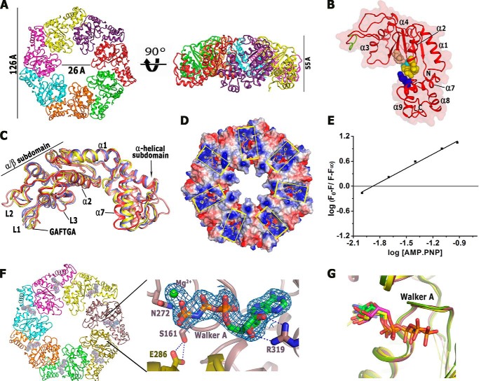
Bacterial enhancer-binding proteins (bEBPs) oligomerize through AAA(+) domains and use ATP hydrolysis-driven energy to isomerize the RNA polymerase-σ(54) complex during transcriptional initiation. Here, we describe the first structure of the central AAA(+) domain of the flagellar regulatory protein FlrC (FlrC(C)), a bEBP that controls flagellar synthesis in Vibrio cholerae. Our results showed that FlrC(C) forms heptamer both in nucleotide (Nt)-free and -bound states without ATP-dependent subunit remodeling. Unlike the bEBPs such as NtrC1 or PspF, a novel cis-mediated "all or none" ATP binding occurs in the heptameric FlrC(C), because constriction at the ATPase site, caused by loop L3 and helix α7, restricts the proximity of the trans-protomer required for Nt binding. A unique "closed to open" movement of Walker A, assisted by trans-acting "Glu switch" Glu-286, facilitates ATP binding and hydrolysis. Fluorescence quenching and ATPase assays on FlrC(C) and mutants revealed that although Arg-349 of sensor II, positioned by trans-acting Glu-286 and Tyr-290, acts as a key residue to bind and hydrolyze ATP, Arg-319 of α7 anchors ribose and controls the rate of ATP hydrolysis by retarding the expulsion of ADP. Heptameric state of FlrC(C) is restored in solution even with the transition state mimicking ADP·AlF3. Structural results and pulldown assays indicated that L3 renders an in-built geometry to L1 and L2 causing σ(54)-FlrC(C) interaction independent of Nt binding. Collectively, our results underscore a novel mechanism of ATP binding and σ(54) interaction that strives to understand the transcriptional mechanism of the bEBPs, which probably interact directly with the RNA polymerase-σ(54) complex without DNA looping.

Keywords: ATPase; ATPases Associated with Diverse Cellular Activities (AAA); Bacterial Transcription; Fluorescence; X-ray Crystallography.


Figures

FIGURE 1.
FIGURE 1.
Sequence alignment of AAA+ domains of bEBPs. Sequence alignment of AAA+ domain of FlrC from V. cholerae, NtrC1 from A. aeolicus, NtrC4 from A. aeolicus, ZraR from S. typhimurium, PspF from E. coli, and dicarboxylic acid transport protein D (DctD) from R. meliloti. Numbering is based on FlrC sequence. Important motifs are indicated by black bars and/or marked. A few important residues are marked by asterisks.
FIGURE 2.
FIGURE 2.
Overall structure of monomer and heptamer of FlrCC in Nt-free and AMP-PNP-bound states. A, at left is a top view illustrating how the protomers pack to form the heptamer of FlrCC; at right is a side view of the same. B, kidney-shaped monomer of FlrCC. GAFTGA motif is colored in green. Walker A (yellow), Walker B (pink), sensor I (cyan), and sensor II (blue) residues are shown as spheres. C, superposition of the Nt-free AAA+ structures of NtrC1 (blue), NtrC4 (yellow), and Pspf (pink) on Nt-free FlrCC (red) showing constriction at the ATP binding pocket of FlrCC, caused by the inclination of α7 and protrusion of L3 loop. D, electrostatic surface of AMP-PNP bound FlrCC showing synchronized binding of AMP-PNP in the ATP binding pockets that are marked by yellow boxes. E, sample plot of fluorescence data from titration of FlrCC with AMP-PNP. F0, F, and F are the relative fluorescence intensities at 338 nm of FlrCC alone, FlrCC in the presence of a given concentration of AMP-PNP, and FlrCC saturated with AMP-PNP, respectively. Slope of the straight line indicates binding stoichiometry. F, at left is 2FoFc electron density map (contoured at 1σ) around AMP-PNP molecules bound to FlrCC heptamer, and at right is the zoomed view of the electron density around an AMP-PNP molecule. G, conformation of Walker A in AMP-PNP-bound FlrCC resembles those of ADP-bound NtrC1C (accession code 1NY6) and ATP-bound NtrC1C (accession code 3M0E). AMP-PNP molecule bound to FlrCC also superposes on ADP and ATP bound to NtrC1C, reflecting canonical binding.
FIGURE 3.
FIGURE 3.
Details of the interactions of AMP-PNP with FlrCC. A, stereo view of 2FoFc electron density map (contoured at 1σ) around the ATP-binding site and bound AMP-PNP. B, hydrophobic packing of the adenine base of AMP-PNP by FlrCC. C, polar interactions to stabilize γ-phosphate and ribose sugar. Interaction of trans-acting Arg-285 with cis-acting Ala-189 of L3 loop is also shown here. D, superposition of AMP-PNP-bound FlrCC (violet) on the Nt-free structure (green) showing structural changes occurred due to AMP-PNP binding. E, interaction of Mg2+ to γ-phosphate and Asp-230. Conserved water Wat1 (W1) and some the other water molecules located close to the γ-phosphate are also shown here. F, superposition of AMP-PNP bound FlrCC (violet) on the Nt-free structure (green) to show the conformational rearrangement of Walker A upon AMP-PNP binding. G, interactions of Walker A before and after AMP-PNP binding.
FIGURE 4.
FIGURE 4.
Trp quenching and ATPase assay with FlrCC and its mutants. A, at left are the plots of ΔFFmax versus AMP-PNP/ADP concentration (in mm) and corresponding Kd values (both in graphical and numerical modes) for FlrCC and its mutants at right. B, ATPase activities of FlrCC ± Mg2+ and the mutants were measured by Malachite green assay and the release of inorganic phosphate (Pi) was estimated against the standard curve of KH2PO4 (left).
FIGURE 5.
FIGURE 5.
Structural details of L1, L2, and L3 loops and interaction of FlrCC with σ54. A, stereo view of 2FoFc electron density map (contoured at 1σ) around L1 loops (in yellow) and L3 loops (in blue) in AMP-PNP-bound FlrCC. B, overall comparison of the structures of Nt-free (gray) and AMP-PNP-bound FlrCC. C, comparisons of the intra-subunit interactions at L1, L2, and L3 loops between Nt-free (gray) and AMP-PNP-bound (magenta) FlrCC. D, inter-protomeric interactions between L1 (magenta) with trans-acting L2 (yellow) and L3 (magenta) with trans-acting α4 (yellow). Nt-free FlrCC is shown in gray. E, comparison of the main chain B-factors for Nt-free (black) and AMP-PNP-bound (red) FlrCC showing an increase of thermal vibration of L1 and L3 loops and a decrease of thermal vibration in α7 and sensor II upon AMP-PNP binding. F, clustering of the high thermal vibration regions (L1 and L3) in AMP-PNP-bound FlrCC (from low, blue, to high, red). G, clustering of the high thermal vibration regions (L1 loops) in ATP-bound NtrC1C(accession code 3M0E) (from low, blue, to high, red). H, pulldown assay showing interactions between FlrCC and σ54 of V. cholerae in the presence and absence of ATP/AMP-PNP. The interaction between FlrCC and σ54 was determined by Coomassie staining. Left gel, lane 1 shows His6-tagged full-length σ54 bound to Ni-NTA resin; lane 2 is the molecular weight marker; lane 3 shows the interaction between FlrCC and His6-tagged σ54 in the absence of Nt; lanes 5 and 7 show the interactions between σ54 and FlrCC in the presence of ATP and AMP-PNP, respectively. Lanes 4, 6, and 8 show background binding of FlrCC with Ni-NTA free and in the presence of ATP and AMP-PNP, respectively. Right gel, purified σ54 (lane 1) and FlrCC (lane 3).
FIGURE 6.
FIGURE 6.
Heptamers of FlrCC in solution, constriction of FlrCC monomer, and comparison with NtrC1C. A, size-exclusion chromatography profiles of FlrCC in the presence or absence of Nt show exclusive formation of the heptamers. The developed chromatograms are shown for FlrCC alone (black), FlrCC + AMP-PNP (blue), and FlrCC + ADP.AlFx (red). The molecular weight of the peaks was determined from the calibration curve prepared using molecular weight standards. B, from left to right, the DLS profiles of Nt-free FlrCC, FlrCC + AMP-PNP, and FlrCC + ADP.AlFx. C, monomer and heptamer of NtrC1C in ATP-bound state. D, monomer and heptamer of FlrCC in AMP-PNP-bound state. ATP binding pocket of the monomers is shown by black bar in C and D.

Similar articles

Cited by

References

    1. Neuwald A. F., Aravind L., Spouge J. L., Koonin E. V. (1999) AAA+: a class of chaperone-like ATPases associated with the assembly, operation, and disassembly of protein complexes. Genome Res. 9, 27–43 - PubMed
    1. Bush M., Dixon R. (2012) The role of bacterial enhancer binding proteins as specialized activators of σ54-dependent transcription. Microbiol. Mol. Biol. Rev. 76, 497–529 - PMC - PubMed
    1. De Carlo S., Chen B., Hoover T. R., Kondrashkina E., Nogales E., Nixon B. T. (2006) The structural basis for regulated assembly and function of the transcriptional activator NtrC. Genes Dev. 20, 1485–1495 - PMC - PubMed
    1. Batchelor J. D., Doucleff M., Lee C. J., Matsubara K., De Carlo S., Heideker J., Lamers M. H., Pelton J. G., Wemmer D. E. (2008) Structure and regulatory mechanism of Aquifex aeolicus NtrC4: variability and evolution in bacterial transcriptional regulation. J. Mol. Biol. 384, 1058–1075 - PubMed
    1. Doucleff M., Chen B., Maris A. E., Wemmer D. E., Kondrashkina E., Nixon B. T. (2005) Negative regulation of AAA+-ATPase assembly by two component receiver domains: a transcription activation mechanism that is conserved in mesophilic and extremely hyperthermophilic bacteria. J. Mol. Biol. 353, 242–255 - PubMed

Publication types

MeSH terms