

doi: 10.1039/c4cc08665a.
Micropatterned, clickable culture substrates enable in situ spatiotemporal control of human PSC-derived neural tissue morphology
Affiliations
- PMID: 25688384
- PMCID: PMC4456773
- DOI: 10.1039/c4cc08665a
Item in Clipboard
Micropatterned, clickable culture substrates enable in situ spatiotemporal control of human PSC-derived neural tissue morphology
Chem Commun (Camb).
.
Abstract
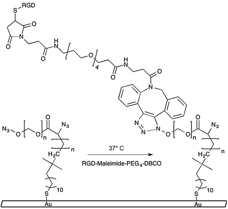
We describe a modular culture platform that enables spatiotemporal control of the morphology of 2D neural tissues derived from human pluripotent stem cells (hPSCs) by simply adding clickable peptides to the media. It should be widely applicable for elucidating how spatiotemporal changes in morphology and substrate biochemistry regulate tissue morphogenesis.
Figures
Similar articles
-
In vitro culture and directed osteogenic differentiation of human pluripotent stem cells on peptides-decorated two-dimensional microenvironment.ACS Appl Mater Interfaces. 2015 Mar 4;7(8):4560-72. doi: 10.1021/acsami.5b00188. Epub 2015 Feb 18. ACS Appl Mater Interfaces. 2015. PMID: 25671246
-
Efficient Expansion of Dissociated Human Pluripotent Stem Cells Using a Synthetic Substrate.Methods Mol Biol. 2016;1307:61-9. doi: 10.1007/7651_2014_82. Methods Mol Biol. 2016. PMID: 24875248
-
Xeno-free adaptation and culture of human pluripotent stem cells.Methods Mol Biol. 2013;1001:81-97. doi: 10.1007/978-1-62703-363-3_8. Methods Mol Biol. 2013. PMID: 23494422
-
The safety of human pluripotent stem cells in clinical treatment.Ann Med. 2015;47(5):370-80. doi: 10.3109/07853890.2015.1051579. Epub 2015 Jul 6. Ann Med. 2015. PMID: 26140342 Review.
-
Microfluidic systems: a new toolbox for pluripotent stem cells.Biotechnol J. 2013 Feb;8(2):180-91. doi: 10.1002/biot.201200206. Epub 2012 Nov 1. Biotechnol J. 2013. PMID: 23125055 Review.
Cited by
-
Synthetic embryology: controlling geometry to model early mammalian development.Curr Opin Genet Dev. 2018 Oct;52:86-91. doi: 10.1016/j.gde.2018.06.006. Epub 2018 Jun 27. Curr Opin Genet Dev. 2018. PMID: 29957587 Free PMC article. Review.
-
Brain Organoids as Model Systems for Genetic Neurodevelopmental Disorders.Front Cell Dev Biol. 2020 Oct 12;8:590119. doi: 10.3389/fcell.2020.590119. eCollection 2020. Front Cell Dev Biol. 2020. PMID: 33154971 Free PMC article. Review.
-
Rethinking developmental toxicity testing: Evolution or revolution?Birth Defects Res. 2018 Jun 1;110(10):840-850. doi: 10.1002/bdr2.1212. Epub 2018 Feb 12. Birth Defects Res. 2018. PMID: 29436169 Free PMC article.
-
High-content imaging with micropatterned multiwell plates reveals influence of cell geometry and cytoskeleton on chromatin dynamics.Biotechnol J. 2015 Oct;10(10):1555-67. doi: 10.1002/biot.201400756. Epub 2015 Jul 14. Biotechnol J. 2015. PMID: 26097126 Free PMC article.
-
In Vitro Microscale Models for Embryogenesis.Adv Biosyst. 2018 Jun;2(6):1700235. doi: 10.1002/adbi.201700235. Epub 2018 May 7. Adv Biosyst. 2018. PMID: 30533517 Free PMC article.
References
Publication types
MeSH terms
Substances
Grants and funding
LinkOut - more resources
Full Text Sources
Other Literature Sources