. 2015 Apr;41(7):925-39.
doi: 10.1111/ejn.12849. Epub 2015 Feb 16.

Spatio-temporal dynamics of adaptation in the human visual system: a high-density electrical mapping study

Affiliations

Spatio-temporal dynamics of adaptation in the human visual system: a high-density electrical mapping study

Gizely N Andrade et al. Eur J Neurosci. 2015 Apr.

Abstract

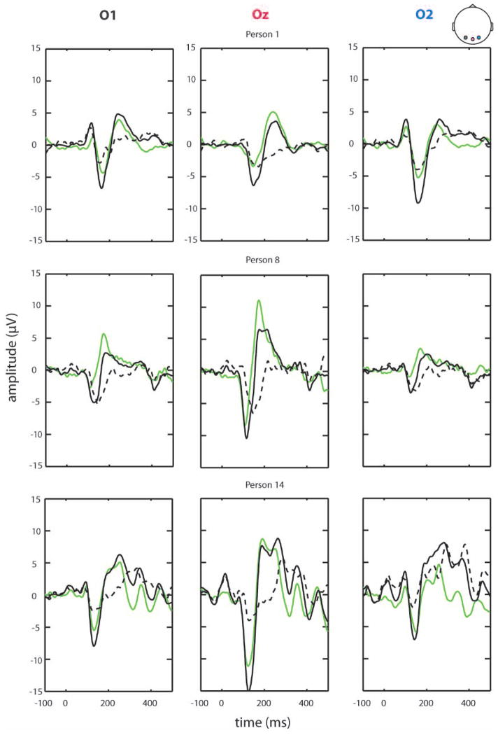
When sensory inputs are presented serially, response amplitudes to stimulus repetitions generally decrease as a function of presentation rate, diminishing rapidly as inter-stimulus intervals (ISIs) fall below 1 s. This 'adaptation' is believed to represent mechanisms by which sensory systems reduce responsivity to consistent environmental inputs, freeing resources to respond to potentially more relevant inputs. While auditory adaptation functions have been relatively well characterized, considerably less is known about visual adaptation in humans. Here, high-density visual-evoked potentials (VEPs) were recorded while two paradigms were used to interrogate visual adaptation. The first presented stimulus pairs with varying ISIs, comparing VEP amplitude to the second stimulus with that of the first (paired-presentation). The second involved blocks of stimulation (N = 100) at various ISIs and comparison of VEP amplitude between blocks of differing ISIs (block-presentation). Robust VEP modulations were evident as a function of presentation rate in the block-paradigm, with strongest modulations in the 130-150 ms and 160-180 ms visual processing phases. In paired-presentations, with ISIs of just 200-300 ms, an enhancement of VEP was evident when comparing S2 with S1, with no significant effect of presentation rate. Importantly, in block-presentations, adaptation effects were statistically robust at the individual participant level. These data suggest that a more taxing block-presentation paradigm is better suited to engage visual adaptation mechanisms than a paired-presentation design. The increased sensitivity of the visual processing metric obtained in the block-paradigm has implications for the examination of visual processing deficits in clinical populations.

Keywords: EEG; habituation; inhibition; plasticity; vision.


Conflict of interest statement

Conflict of Interest Statement

All authors of this paper declare no conflicts-of-interest, financial or otherwise, that could have biased their contributions to this work. The senior author, Dr. Foxe, attests that all authors had access to the full dataset and to all stages of the analyses.

Figures

Figure 1
Figure 1
Schematic diagram illustrating the two paradigms. A. Depiction of the paired-presentation paradigm used in Experiment 1. Stimuli were presented in pairs with an inter-stimulus interval of either 200 or 300ms and a long inter-pair interval of 2500ms. Catch trials, consisting of unpaired checkerboards, were presented one third of the time and were used to extract the isolated response to the second stimulus in a pair. B. Depiction of the block-presentation paradigm used in Experiment 2. Stimuli were presented in blocks of 100 trials, at an inter-stimulus interval centered around ISIs of 200, 300, 550, 1050, or 2550ms. The stimulus presentation was jittered by +/−50ms to allow for the implementation of an ADJAR procedure which models and removes response overlap (used in the 200ms condition).
Figure 2
Figure 2
Waveforms obtained from subtracting the catch trials from the 200 and 300ms trials. The catch serves as the pure response to a single stimulus presentation (S1) and the trial waveform minus the catch represents the ‘isolated’ response to the second stimulus (S2). At 140ms there is an effect of order, with VEPs to the S2 being greater than to the S1. At 170ms, the effect reverses, with VEPs to the S2 being smaller (less positive) than to the S1. However, neither of these effects depends on ISI.
Figure 3
Figure 3
Scalp topographic maps for the paired-paradigm reveal a difference between the first and second stimulus presentations and between the second and the catch for both ISIs. Additionally there is a clear difference in topography when comparing the response at 100 and 140ms (strong negativity) with the response at 170ms (strong positivity).
Figure 4
Figure 4
A. Average waveforms for each of the five ISI conditions (Experiment 2) are displayed for the five occipital and occipito-parietal scalp sites of interest. A clear effect of ISI on VEP amplitude can be seen between 100–200ms, with slower stimulus presentation rates leading to greater absolute VEP amplitude. B. Amplitude by ISI plots. The effect of ISI on VEP amplitude at each scalp site for the three time periods of interest. The greatest adaptation based on ISI is seen in the later timewindows (130–150ms & 160–180ms) and is most robust at the central occipital site. Additionally, the directionality of adaptation by ISI at the central site (Oz) is exclusively reversed in the last time period of interest, with the slowest ISI here eliciting the smallest VEP amplitudes.
Figure 5
Figure 5
Left - Scalp topographic maps reveal similar central-occipital negativity for the early time period across ISIs in the block-presentation. At the later time points the voltage distribution diverges, with the shorter ISIs showing central-occipital positivity and the longer ISIs a bilateral negativity. Right- Scalp topographic maps depicting the difference in electrical activation when comparing the 2550ms condition to each of the other ISIs. The difference topography is most unique when comparing electrical activation across the scalp when comparing the 1050ms ISI to the 2050ms ISI at 140 & 180ms post stimulus presentation.
Figure 6
Figure 6
A. Brain generators estimated using a distributed linear inverse solution on the local autoregressive average for block-design. Early sources are mainly over occipital cortex, with later sources extending over parietal and temporal cortex. B. Randomization tests reveal significant differences in brain generators between the 300ms and 2550ms conditions. At 90–110ms these differences are mostly occipital. At 130–150ms the differences are most pronounced and expand over occipital, parietal, temporal and frontal areas, including superior parietal cortex and lateral occipital cortex. At 160–180ms, the only significantly different sources are frontal.
Figure 7
Figure 7
Representative individual participant visual evoked potential waveforms. VEP to a ‘fast’ ISI condition (300ms) and ‘slow’ ISI condition (2550ms) is plotted for the central occipital site. The dashed line represents the difference in amplitude between these two conditions and can be interpreted as an index of adaptation.

Similar articles

Cited by

References

    1. Adler LEW, MC, Freedman R. Neurophysiologic Studies of Sensory Gating in Schizophrenia: Comparison of Auditory and Visual Responses. Biol Psychiatry. 1985;20:1284–1296. - PubMed
    1. Arnfred SM, Eder DN, et al. Gating of the vertex somatosensory and auditory evoked potential P50 and the correlation to skin conductance orienting response in healthy men. Psychiatry Res. 2001;101(3):221–235. - PubMed
    1. Bedwell JS, Chan CC, et al. Changes in the visual-evoked P1 potential as a function of schizotypy and background color in healthy young adults. J Psychiatr Res. 2013;47(4):542–547. - PubMed
    1. Braff DL, Light GA, et al. Prepulse inhibition and P50 suppression are both deficient but not correlated in schizophrenia patients. Biol Psychiatry. 2007;61(10):1204–1207. - PubMed
    1. Brockhaus-Dumke A, Mueller R, et al. Sensory gating revisited: Relation between brain oscillations and auditory evoked potentials in schizophrenia. Schizophrenia Research. 2008;99(1–3):238–249. - PubMed

Publication types

LinkOut - more resources