. 2015 Feb 17;13(2):e1002069.
doi: 10.1371/journal.pbio.1002069. eCollection 2015 Feb.

Luminal progenitors restrict their lineage potential during mammary gland development

Affiliations

Luminal progenitors restrict their lineage potential during mammary gland development

Veronica Rodilla et al. PLoS Biol. .

Abstract

The hierarchical relationships between stem cells and progenitors that guide mammary gland morphogenesis are still poorly defined. While multipotent basal stem cells have been found within the myoepithelial compartment, the in vivo lineage potential of luminal progenitors is unclear. Here we used the expression of the Notch1 receptor, previously implicated in mammary gland development and tumorigenesis, to elucidate the hierarchical organization of mammary stem/progenitor cells by lineage tracing. We found that Notch1 expression identifies multipotent stem cells in the embryonic mammary bud, which progressively restrict their lineage potential during mammary ductal morphogenesis to exclusively generate an ERαneg luminal lineage postnatally. Importantly, our results show that Notch1-labelled cells represent the alveolar progenitors that expand during pregnancy and survive multiple successive involutions. This study reveals that postnatal luminal epithelial cells derive from distinct self-sustained lineages that may represent the cells of origin of different breast cancer subtypes.


Conflict of interest statement

The authors have declared that no competing interests exist.

Figures
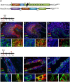
Fig 1
Fig 1. Notch1 is expressed in multipotent embryonic stem cells.
(A) Schematic representation of the Notch1-CreERT2SAT knock-in mice (referred to as N1CreERT2) and Rosa26mTomato/mGFP reporter mice (called R26mTmG) used in this study. Pregnant females were induced with tamoxifen to label their embryos at embryonic day E15.5 and double transgenic N1CreERT2R26mTmG littermates were analyzed 24 h later (B–D) or 5 wk after birth (E–G). (B–D) Representative embryonic mammary bud sections show that Notch1pos cells (marked by GFP in green) express both myoepithelial (K5, in red in B) and luminal markers (K8, in red in C) and they are negative for ERα (in red in D), n = 2. (E–G) Representative pubertal mammary gland sections show that Notch1-derived clones (in green) contain myoepithelial (K5pos in red in E) and luminal cells (K8pos in red in F) as well as ERαpos and ERαneg cells (ERα in red in G), n = 3. 4',6-diamidino-2-phenylindole (DAPI) stains DNA in blue. Scale bars correspond to 20 µm in B–G, and 10 µm in the magnifications of the insets.
Fig 2
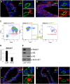
Fig 2. Notch1 expression in the postnatal mammary gland is restricted to ERαneg and PRneg luminal cells.
(A–B) Representative sections of ducts from pubertal (6-wk-old) N1CreERT2R26mTmG females analyzed 24 h upon tamoxifen injection. Immunofluorescence was performed with anti-K5 antibodies (in red in A), anti-K8 (in red in B), anti-GFP (to reveal Notch1-marked cells in green) and DAPI stains DNA in blue; n = 3. (C) FACS plots of 6-wk-old N1CreERT2R26mTmG females analyzed 24 h upon tamoxifen injection. Dissociated mammary cells were gated as Linneg cells (CD45/CD31/Ter119)neg, and then as mammary epithelial cells (MEC) using the CD24 and CD29 markers, allowing us to resolve luminal (CD24+CD29low) and myoepithelial (CD24+CD29high) populations. 98.24 ±0.4% of GFPpos cells gated in MEC were found in the luminal subset by FACS analysis. Note that GFPpos cells also display Tomato fluorescence 24 h after induction, as the Tomato protein is still present at this time point, even if recombination has occurred. Values are shown in average ± s.e.m, n = 6. (D–E) The expression of Notch1 in sorted luminal (CD24+CD29low) (LUM) and myoepithelial cells (CD24+CD29high) (MYO) mammary cells from 10-wk-old B6/N wild-type females was analyzed at the mRNA level by qRT-PCR in A and at the protein level by western blot in B. The relative mRNA expression was normalized to the housekeeping gene 18S in A, while lamin B1 was used as a loading control in E. For western blot analysis, we used anti-K8 and anti-K5 antibodies as controls for sorted luminal and myoepithelial cells, respectively. n = 2. (F–G) Representative sections of mammary ducts from 6-wk-old N1CreERT2R26mTmG females show that GFP-expressing cells (in green) are invariably negative for ERα (in red in F) and PR expression (in red in G). DAPI stains DNA in blue, n = 3. Scale bars correspond to 20 µm in C–D and F–G and 10 µm in the insets.
Fig 3
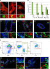
Fig 3. ERαneg luminal cells represent self-sustained mammary progenitors that clonally expand and self-renew in vivo.
(A–B) N1CreERT2R26mTmG females induced with a low dose of tamoxifen (1µg/g of mouse body weight) at 4 wk of age and analyzed at different time points: 24 h (pulse), 2 wk (2 wk chase), 6 wk (6 wk chase), or 10 wk later (10 wk chase). (A) Representative images of whole mount digestion of mammary ducts labeling single cells (one cell), small clones (two to five cells), and large clones (six or more cells). Notch1-labelled cells are in green and Tomato fluorescence labels non-targeted cells. n = 11 mice. (B) Quantification of the in vivo clonal expansion of GFP-marked cells, at the indicated time points; n = 11. (C–E) N1CreERT2R26mTmG females were induced with a single dose of tamoxifen (0.1mg/g of mouse body weight) at 4 wk of age and analyzed 10 wk later. The clonal progeny of Notch1-expressing cells (marked in green by GFP) is still luminal (K5neg in red in C), and ERαneg (in red in D) and PRneg (in red in E), as the cells from which it derives. DAPI stains DNA in blue, n = 4. (F) FACS plots of N1CreERT2R26mTmG females induced at puberty and analyzed after three cycles of pregnancy, lactation, and involution. Mammary epithelial cells (MEC) from females in the third week of the last involution were dissociated and analyzed by flow cytometry. GFPpos cells represent 28.9 ±1.4% of the luminal population (CD24+and CD29low), n = 2. (G–H) Representative sections of mammary ducts from N1CreERT2R26mTmG females induced at 4 wk of age and analyzed after three serial pregnancies. GFP-expressing cells (in green) remain invariably negative for ERα (in red in G) and PR expression (in red in H). DAPI stains DNA in blue, n = 2. Scale bars correspond to 40 µm in A; 20 µm in C–E, G and H; and 10 µm in the insets in C–E and G–H.
Fig 4
Fig 4. Notch1-expressing cells define highly clonogenic ERαneg luminal progenitors.
(A) N1CreERT2R26mTmG females induced with tamoxifen at puberty (6 wk of age) and analyzed 24 h later present a high proportion of proliferative GFPpos cells (73.45 ±3.72%), as indicated by Ki67 expression (in red); DAPI stains DNA in blue, n = 5. (B) Left, histogram of the cell cycle profile of total luminal cells (Luminal, in blue) compared to Notch1-expressing cells (GFPpos, in green) sorted by FACS and stained with Hoechst-33342 to evaluate their DNA content. Right, quantification of cycling (S/G2/M) and non-cycling (G0/G1) cells in total luminal cells (blue, S/G2/M = 26.9 ±5.5%) and in Notch1-expressing cells (green, S/G2/M = 47.0 ±5.2%). Data are represented as a mean ± s.e.m. of n = 5 animals, (*) p < 0.05 with t test. (C) N1CreERT2R26mTmG females were induced with tamoxifen at 10 wk of age and analyzed 24 h later. GFPneg and GFPpos cells were sorted as Lin-CD24+CD29low luminal cells by FACS, seeded on a feeder layer of irradiated fibroblast and their clonogenic capacity was evaluated after 7 d in culture. Top, quantification of the number of colonies generated by GFPneg and GFPpos cells. n = 3 experiments with two mice each, (***) p < 0.001 with t test. Bottom, representative pictures of counted colonies stained with Hematoxylin are shown below each bar. (D) Flow cytometry analysis indicates that GFPpos cells (in green) are found within progenitor cell populations (CD49bpos, Sca1neg and CD133neg) gated in the luminal population (Lin-CD24+CD29low) (in blue). GFPpos cells represent 82.02 ±3.28% of CD49bpos cells, 96.9 ±0.6% of Sca1neg cells, and 93.7 ±1.0% of CD133neg cells. n = 10. Scale bars correspond to 20 µm in A and 10 µm in the insets in A.
Fig 5
Fig 5. The transcriptional signature of ERαneg mammary luminal progenitors is conserved in their derived lineages.
(A) Genome-wide heat map showing the pattern of GFPneg and GFPpos luminal cells (Lin-CD24+CD29low) from 10-wk-old N1CreERT2R26mTmG females induced for 24 h. Biological triplicates for each group were performed by pooling two females per experiment and are represented by different columns in the unsupervised clustering. (B) GSEA analysis showing the enrichment plots for GFPneg and GFPpos transcriptome profiles in four published gene signatures (Luminal_Mature_Up, Luminal_Mature_Down, Luminal_Progenitor_Up, Luminal_ Progenitor_Down). Sorted GFPneg cells correlate with luminal mature cells, while sorted GFPpos cells correspond to luminal progenitors. ES: Enrichment Score. (C) qRT-PCR analysis, in adult N1CreERT2R26mTmG females induced for 24 h, of the top-ten ranked genes in the transcriptomic analysis (first row: up-regulated in GFPpos and second row: down-regulated in GFPpos) confirms the differential expression of each of these genes detected in the microarray experiments. The expression levels of each gene in sorted myoepithelial cells are also shown (Myo). Relative mRNA expression was normalized to the housekeeping gene 18S. (*) p < 0.05, (**) p < 0.01, (***) p < 0.001 with t test. n = 2. (D) FACS analysis of sorted cells from N1CreERT2R26mTmG females induced at 4 wk of age and analyzed 10 wk later (n = 8) shows that GFPpos cells (Notch1-derived progeny, in green) overlap with ERneg luminal progenitor markers (CD49bpos, Sca1neg and CD133neg in blue) gated as Lin-CD24+CD29low luminal cells, showing an identical profile as Notch1-expressing cells 24 h post-induction (see Fig. 4D).
Fig 6
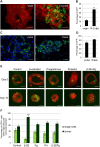
Fig 6. Notch1-expressing cells contribute to alveologenesis at pregnancy and expand in response to hormonal stimulation.
(A) N1CreERT2R26mTmG females were induced with a low dose of tamoxifen (1µg/g of mouse body weight) at 4 wk of age and analyzed as virgins (left) or at mid-pregnancy (14.5 d post coitum, dpc) (right panel). Representative pictures of whole mount mammary gland ducts are shown, where red denotes tomato fluorescence, while green indicates Notch1-derived GFPpos cells. n = 3. (B) Quantification by flow cytometry of the percentage of GFPpos luminal cells (Lin-CD24+CD29low) from N1CreERT2R26mTmG females induced at 4 wk of age (0.1mg/g of mouse body weight) and analyzed at virgin or pregnant state (14.5 dpc). An extensive increase in the amount of GFPpos cells is evident at pregnancy in A and B. n = 7. (**) p = 0.001 with t test. (C) Left, representative mammary section of N1CreERT2R26mTmG females injected with tamoxifen at mid-pregnancy (14.5 dpc) and analyzed 24 h later (pulse). Right, mammary section of N1CreERT2R26mTmG female injected at 4 wk of age and analyzed at mid-pregnancy (chase). (D) Quantification by flow cytometry of the percentage of GFPpos cells luminal cells (Lin-CD24+CD29low) from N1CreERT2R26mTmG females injected at mid-pregnancy (pulse: 69.75 ±3.55%) compared to females injected at 4 wk of age and analyzed at mid-pregnancy (chase: 86.1 ±2.01%). Values are shown as percentages ±s.e.m. of n = 5. (E) 3-D organotypic cultures from adult N1CreERT2R26mTmG females were induced with 4-hydroxytamoxifen (4-OHT) in vitro and treated for 10 d with different hormones, as indicated. Red denotes tomato fluorescence and green indicates Notch1-marked cells at Day 0 and progeny at Day 10; n = 4. (F) Quantification of the clonal expansion of GFPpos cells in 3-D organoids, classified as single cells (one cell, in light green) or clones (two or more cells, in dark green) indicates a significant expansion only in response to estradiol (β-2E bars). (***) p < 0.001 with t test. Scale bars correspond to 40 µm in A and E and 20 µm in C.
Fig 7
Fig 7. Notch1-expressing cells reveal a high regenerative capacity in transplantation assays.
(A) 10,000 GFPpos luminal cells were sorted by FACS within the CD24+/CD29low gate, mixed 1:1 with Tomato epithelial cells (Linneg), and injected in cleared fat pads of host mice. All transplanted animals showed outgrowths (n = 2). GFPpos cells generated luminal cells but not myoepithelial cells, stained with anti-K5 antibody (in red in A). (B–C) Whole-mounts of GFPpos outgrowths obtained in virgin host mice (n = 1 out of 7 transplanted, in B) and pregnant host females (n = 7 out of 11 transplanted, in C). On the right of each panel, graphic representations of the extent to which each transplant filled the fat pad (in black). (D–F) 10,000 sorted luminal GFPpos (Notch1-expressing cells) cells were injected per fat pad, in the absence of myoepithelial cells. Immunofluorescence of representative sections of obtained outgrowths show that GFPpos cells generate all mammary lineages, both in virgin (D–E) and in pregnant recipient mice (F). Immunofluorescence labeling of myoepithelial cells (anti-K5 in red in D), and ERαpos (in red in E and F) show that GFP-derived clones contain K5pos myoepithelial cells, ERαpos and ERαneg luminal cells. Scale bars correspond to 10 µm in A, 2 mm in B–C, and 20 µm in D–F.
Fig 8
Fig 8. Proposed model for luminal cell hierarchy during mammary gland development.
(A) Lineage tracing from embryos reveals that Notch1-expressing (N1pos) multipotent stem cells exist only during embryonic mammary development, when they co-express myoepithelial cytokeratin (K5) and luminal cytokeratin (K8). These multipotent stem cells can generate all mammary lineages (labeled in green postnatally). (B) After birth, luminal stem cells resolve in two distinct luminal progenitors: ERαpos and ERαneg cells, which maintain exclusively their own lineage throughout adulthood. Unipotent progenitors lacking ERα expression (ERαneg) marked by Notch1 are responsible for generating milk-producing alveoli at pregnancy and define self-renewing luminal cells able to survive mammary gland involution.

References

    1. Shackleton M, Vaillant F, Simpson KJ, Stingl J, Smyth GK, et al. (2006) Generation of a functional mammary gland from a single stem cell. Nature 439: 84–88. - PubMed
    1. Stingl J, Eirew P, Ricketson I, Shackleton M, Vaillant F, et al. (2006) Purification and unique properties of mammary epithelial stem cells. Nature 439: 993–997. - PubMed
    1. Rios AC, Fu NY, Lindeman GJ, Visvader JE (2014) In situ identification of bipotent stem cells in the mammary gland. Nature 506: 322–327. 10.1038/nature12948 - DOI - PubMed
    1. Van Keymeulen A, Rocha AS, Ousset M, Beck B, Bouvencourt G, et al. (2011) Distinct stem cells contribute to mammary gland development and maintenance. Nature 479: 189–193. 10.1038/nature10573 - DOI - PubMed
    1. Molyneux G, Geyer FC, Magnay FA, McCarthy A, Kendrick H, et al. (2010) BRCA1 basal-like breast cancers originate from luminal epithelial progenitors and not from basal stem cells. Cell Stem Cell 7: 403–417. 10.1016/j.stem.2010.07.010 - DOI - PubMed

Publication types

MeSH terms

LinkOut - more resources