. 2015 Feb 17;10(2):e0117444.
doi: 10.1371/journal.pone.0117444. eCollection 2015.

Investigating the role of RIO protein kinases in Caenorhabditis elegans

Affiliations

Investigating the role of RIO protein kinases in Caenorhabditis elegans

Tasha K Mendes et al. PLoS One. .

Erratum in

Abstract

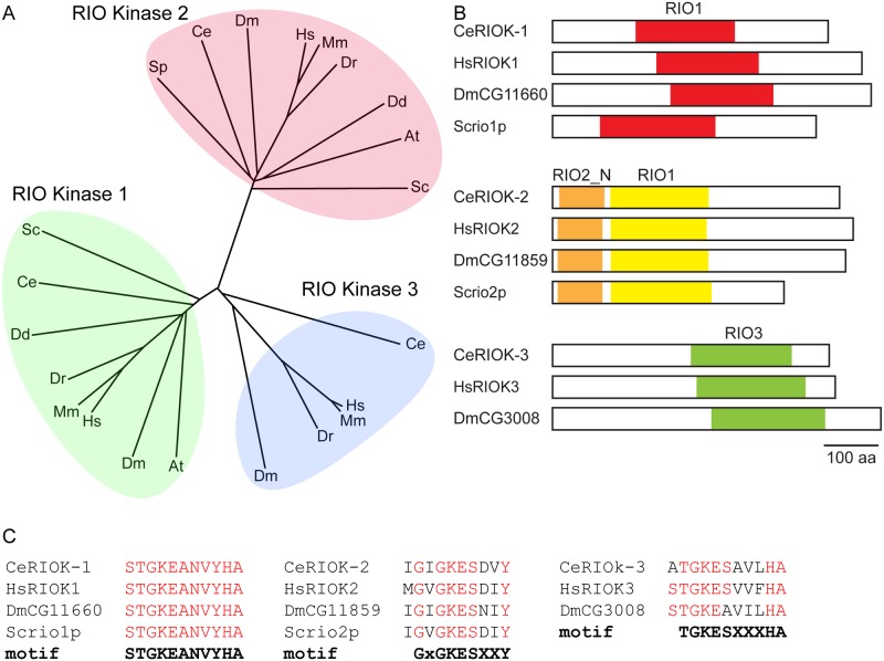
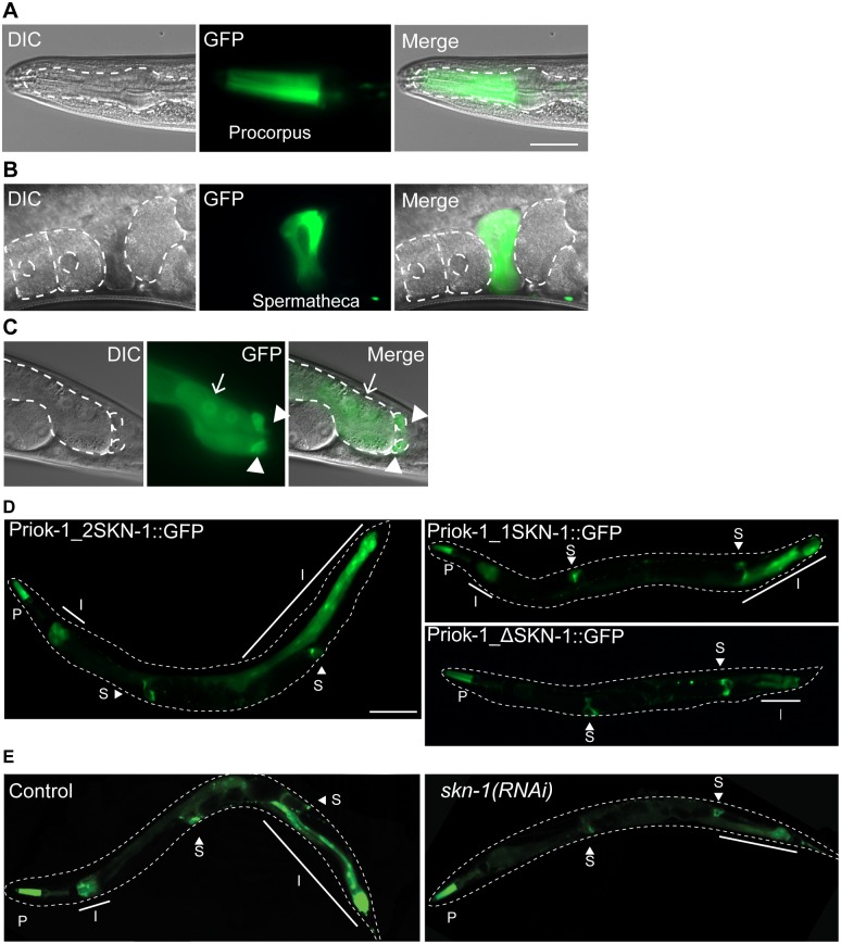
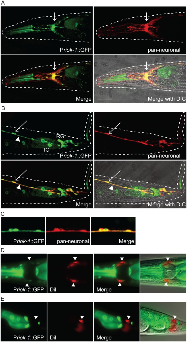
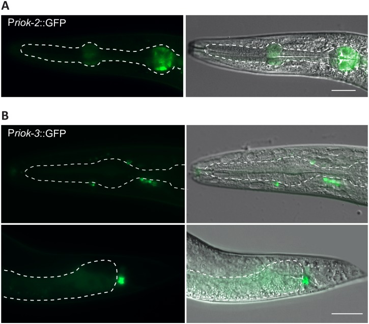
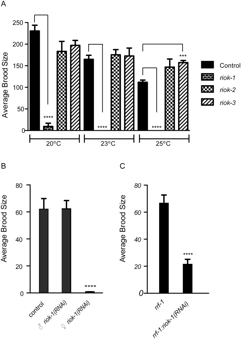
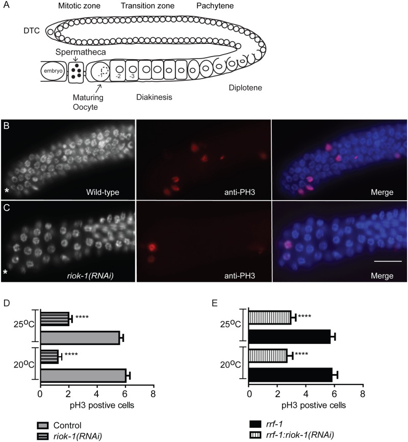
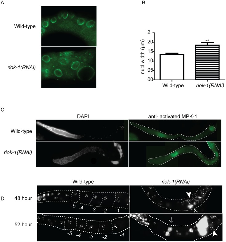
RIO protein kinases (RIOKs) are a relatively conserved family of enzymes implicated in cell cycle control and ribosomal RNA processing. Despite their functional importance, they remain a poorly understood group of kinases in multicellular organisms. Here, we show that the C. elegans genome contains one member of each of the three RIOK sub-families and that each of the genes coding for them has a unique tissue expression pattern. Our analysis showed that the gene encoding RIOK-1 (riok-1) was broadly and strongly expressed. Interestingly, the intestinal expression of riok-1 was dependent upon two putative binding sites for the oxidative and xenobiotic stress response transcription factor SKN-1. RNA interference (RNAi)-mediated knock down of riok-1 resulted in germline defects, including defects in germ line stem cell proliferation, oocyte maturation and the production of endomitotic oocytes. Taken together, our findings indicate new functions for RIOK-1 in post mitotic tissues and in reproduction.


Conflict of interest statement

Competing Interests: The authors have declared that no competing interests exist.

Figures

Fig 1
Fig 1. The RIO family of kinases are evolutionary conserved.
(A) Unrooted phylogenetic tree of RIO proteins derived from a Clustal Omega alignment of whole proteins sequences (European Bioinformatics Institute). Tree was drawn using Philodendron Phylogenetic tree printer. Hs, H. sapiens; Mm, M. musculus; Dm, melanogaster; Ce, C. elegans; Xl, X. Laevis; Dr, D. reri; Dd, D. discoideum; At, A. thaliana; Sc, S. cerevisiae (B) Schematic representing the domain organisation in RIO kinases. Each of the RIO kinase sub-families has a conserved domain organisation; Hs, H. sapiens; Dm, melanogaster; Ce, C. elegans; Sc, S. cerevisiae examined. (C) Alignment of the ATP binding loop in the RIO domain.
Fig 2
Fig 2. The putative riok-1 promoter drives GFP expression in multiple tissues.
(A) procorpus region of the pharynx; pharynx is outlined (B) spermatheca; oocytes and eggs are outlined. (C) intestine (arrow) and the rectal gland (arrow heads); intestinal cells and rectal gland outlined. Scale bar = 25μm. (D) Intestinal expression is largely SKN-1 dependent. Representative images of GFP expression in Priok-1_2SKN-1, Priok-1_1SKN-1 and Priok-1_ΔSKN-1 worms. I, intestine; P, Pharynx; S, spermatheca. (E) SKN-1 is required for normal expression of riok-1 in the intestine. RNAi knockdown of skn-1, but not control (pCB19), leads to a reduction in the Priok-1_2SKN-1 intestinal expression. Worms are outlined with a dashed line and the solid line indicates the extent of GFP expression in the intestine. Scale bar = 100 μm.
Fig 3
Fig 3. Priok-1_2SKN-1 drives GFP expression in a small subset of neurons.
(A–C) Single plane confocal images from adult worms. (A) GFP was expressed in a subset of head neurons and is highly expressed in the nerve ring displaying strong co-localisation with the pan-neuronal marker (arrow). (B) Co-localisation of GFP with pan-neuronal dsRED. D-type neurons (arrow) and longitudinal nerves (arrow head). Intestinal cells (IC) and the rectal gland (RG) can also be observed. (C) Zoomed in image of the D-type neurons displaying co-localisation. Scale bar = 25μm. (D–E) Priok-1_2SKN-1 GFP expression does not co-localise with the lipophilic dye Dil, in head neurons (D) or tail neurons (E). Scale bar = 15 μm.
Fig 4
Fig 4. The putative promoters of riok-2 and -3 drive GFP in distinct tissues.
(A) riok-2 transgenic worms displayed weak GFP expression in the metacorpus and posterior bulb of the pharynx. Scale bar = 25μm (B) riok-3 transgenic worms revealed weak GFP expression, in some head and was strongly expressed in a tail neuron which may be the PVQ, PHAL or PQR neuron. Scale bar = 25μm.
Fig 5
Fig 5. riok-1(RNAi) sterility is associated with an oogenesis defect.
(A) Total average brood size for wild-type worms knocked down for riok-1, riok-2 or riok-3 at 20°C, 23°C and 25°C. riok-1(RNAi) worms have a significant reduction in brood size at 20°C and are sterile at 23°C and 25°C. Error bars represent the SEM; n6. **** = P values <0.0001. *** = P values 0.0003 for riok-3 at 25°C (B) Average number of progeny produced in 17 hours from crossing fog-2(q71) male and female worms. Crosses were; control, ♀ fog-2(q71) x ♂ fog-2(q71); ♀ fog-2(q71) x ♂riok-1(RNAi); ♀ fog-2(q71)riok-1(RNAi) x ♂ fog-2(q71). Thirteen individual crosses per treatment were conducted at 25°C with 10 males mated per L4 female. **** = P <0.0001. (C) Knockdown of riok-1 in rrf-1 worms resulted in significantly reduced progeny. Total brood size for rrf-1 worms grown on riok-1 and pCB19 (negative control) RNAi at 25°C. Error bars represent SEM. n = 23, **** = P <0.0001.
Fig 6
Fig 6. riok-1 is required for normal germ cell proliferation.
(A) Overview of the adult C. elegans hermaphrodite gonad. One of the two gonad U-shaped tubular gonad is shown. The single somatic distal tip cell (DTC) maintains a population of self-renewing germline stem cells. Germ cells enter into the meiotic prophase I in the transition zone and then progress to the pachytene stage. During these stages, germ cells are only partially enclosed in a membrane and share a common cytoplasmic core. As germ cells exit pachytene, they move through the “loop” region and enter diplotene and begin to fully cellularise. A signal from sperm directs the most proximal oocytes to undergo maturation and fertilisation occurs are the oocyte moves through the spermatheca into the uterus. (B-C) Dissected gonads were stained with anti-pH3 staining to detect proliferating germ cells and DAPI to visualise DNA. Asterisks indicate the distal end of the gonad. Scale bar = 15μm. (D-E) riok-1(RNAi) gonads had a reduced number of pH3 stained cells in the mitotic zone (D) Wild-type worms with riok-1 knocked down at 20°C and 25°C (n = 50) (E)) rrf-1;pCB19(RNAi) and rrf-1:riok-1(RNAi) worms at 20°C and 25°C (n = 50). Error bars represent SEM; **** = P values <0.0001.
Fig 7
Fig 7. Proximal gonad organization requires riok-1.
(A) Representative images of wild-type and riok-1(RNAi) dissected gonads immunostained stained for nuclear pores. (B) The nuclear diameter of germ cells in the proximal loop region of riok-1(RNAi) worms are significantly increased in size (n = 12). Error bars represent SEM; ** = P values 0.0045. (C) Wild-type and riok-1(RNAi) dissected gonads immunostained stained for activated MAPK. Increased MAPK staining is seen in pachytene and throughout diplotene. (D) Progression of the riok-1(RNAi) EMO phenotype was examined at time points 48 and 52 hours post L1. Gonads from dissected wild-type and riok-1(RNAi) worms were stained with DAPI to visualise DNA. The gonad and oocytes are outlined and proximal wild-type oocytes have been outlined and labelled (-1 to-5). At all time points wild-type worms displayed the typical six bivalent chromosomes in their maturing oocytes within the proximal gonad (n = 30). riok-1(RNAi) worms (n = 30) showed DNA aggregates (arrow) in the proximal region of the gonad and abnormal localised sperm (arrow head). Scale bar = 25 μm; worms grown at 25°C.

References

    1. Manning G, Whyte DB, Martinez R, Hunter T, Sudarsanam S (2002) The protein kinase complement of the human genome. Science 298: 1912–1934. - PubMed
    1. Manning G (2005) Genomic overview of Protein Kinases (December 13, 2005), WormBook, ed. The C. elegans Research Community, WormBook, Available: http://www.wormbook.org. - PMC - PubMed
    1. Hanks SK, Hunter T (1995) Protein kinases 6. The eukaryotic protein kinase superfamily: kinase (catalytic) domain structure and classification. FASEB J 9: 576–596. - PubMed
    1. Scheeff ED, Bourne PE (2005) Structural evolution of the protein kinase-like superfamily. PLOS Comput Biol 1: e49 - PMC - PubMed
    1. Vanrobays E, Gleizes PE, Bousquet-Antonelli C, Noaillac-Depeyre J, Caizergues-Ferrer M, et al. (2001) Processing of 20S pre-rRNA to 18S ribosomal RNA in yeast requires Rrp10p, an essential non-ribosomal cytoplasmic protein. EMBO J 20: 4204–4213. - PMC - PubMed

Publication types

MeSH terms

LinkOut - more resources