. 2015 Mar 18;6(3):494-504.
doi: 10.1021/acschemneuro.5b00022. Epub 2015 Mar 5.

Delivery and tracking of quantum dot peptide bioconjugates in an intact developing avian brain

Affiliations

Delivery and tracking of quantum dot peptide bioconjugates in an intact developing avian brain

Rishabh Agarwal et al. ACS Chem Neurosci. .

Abstract

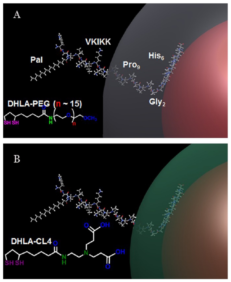
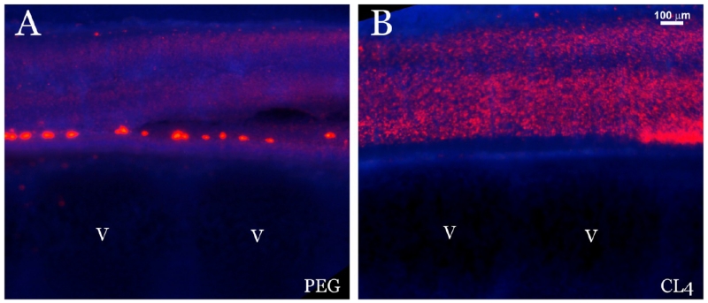
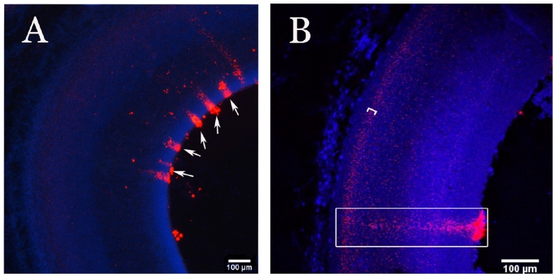
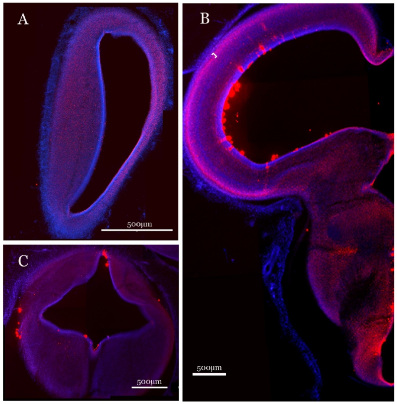
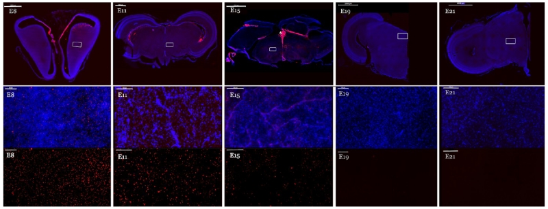
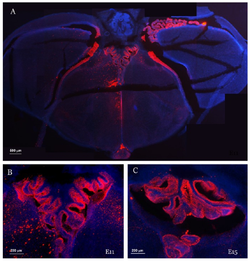
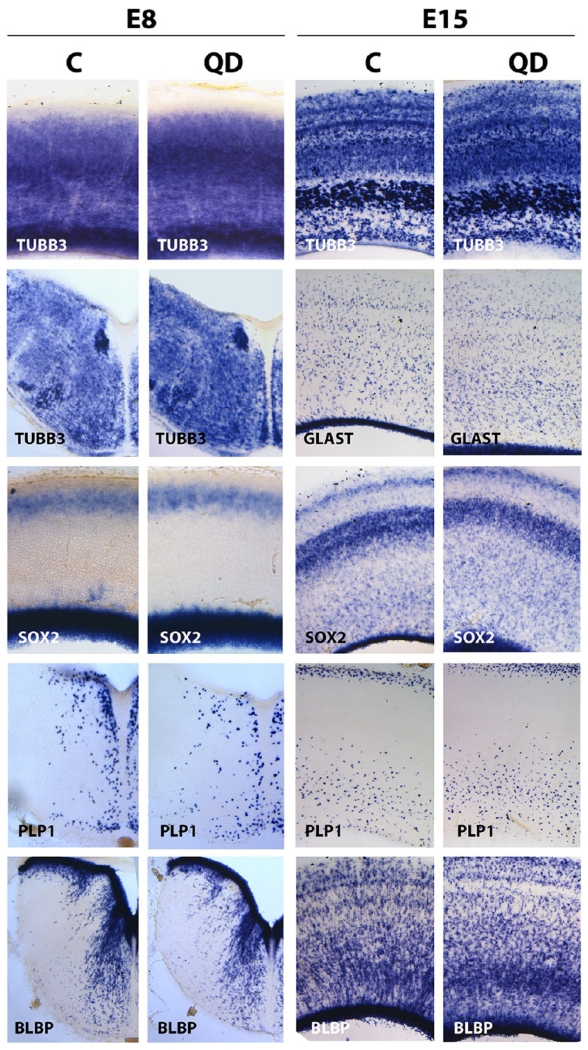
Luminescent semiconductor ∼9.5 nm nanoparticles (quantum dots: QDs) have intrinsic physiochemical and optical properties which enable us to begin to understand the mechanisms of nanoparticle mediated chemical/drug delivery. Here, we demonstrate the ability of CdSe/ZnS core/shell QDs surface functionalized with a zwitterionic compact ligand to deliver a cell-penetrating lipopeptide to the developing chick embryo brain without any apparent toxicity. Functionalized QDs were conjugated to the palmitoylated peptide WGDap(Palmitoyl)VKIKKP9GGH6, previously shown to uniquely facilitate endosomal escape, and microinjected into the embryonic chick spinal cord canal at embryo day 4 (E4). We were subsequently able to follow the labeling of spinal cord extension into the ventricles, migratory neuroblasts, maturing brain cells, and complex structures such as the choroid plexus. QD intensity extended throughout the brain, and peaked between E8 and E11 when fluorescence was concentrated in the choroid plexus before declining to hatching (E21/P0). We observed no abnormalities in embryonic patterning or embryo survival, and mRNA in situ hybridization confirmed that, at key developmental stages, the expression pattern of genes associated with different brain cell types (brain lipid binding protein, Sox-2, proteolipid protein and Class III-β-Tubulin) all showed a normal labeling pattern and intensity. Our findings suggest that we can use chemically modified QDs to identify and track neural stem cells as they migrate, that the choroid plexus clears these injected QDs/nanoparticles from the brain after E15, and that they can deliver drugs and peptides to the developing brain.

Keywords: Nanoparticles; choroid plexus; normal embryonic chick brain development; peptidyl delivery; quantum dots; zwitterion.


Figures

Figure 1
Figure 1
Schematic depicting the QD-peptide structure. (A) Schematic representation of 625 nm QDs surface functionalized with DHLA-PEG (QD-PEG-JB577) and displaying the palmitoylated peptide (JB577) assembled by polyhistidine linkage. The 625 nm QD (diameter ~10 nm) is shown in red, and the PEG as a gray shell extending 30 Å from the QD surface. The peptide is shown in a ball and stick structure appended onto the QD by the hexahistidine sequence. Functional residues along with the position of the palmitate (Pal) group are indicated as well. The Gly2-Pro9 portion of the peptide is shown as almost completely enveloped in the PEG layer. (B) Schematic representation of the 625 QD surface functionalized with DHLA-CL4 (green shell) and binding the same peptide (QD-CL4-JB577). The structures for each respective ligand are shown inset.
Figure 2
Figure 2
Comparison of the fate of QD-PEG-JB577 and QD-CL4-JB577 microinjected into in vivo embryos at E4. Representative sagittal 40 μm section through the spinal cord of E6 embryos injected at E4. (A) Injected with QD-PEG-JB577 peptide conjugate. (B) Injected with QD-CL4-JB577 peptide conjugate. QDs are red and DAPI stained nuclei are blue. A more uniform dispersion of the QDs with the CL4 coating is observed compared to PEG. The position of the vertebrae is indicated (V). The results presented here and in the following images are representative of 5 independent injection experiments each involving 18 control and 18 QD-injected embryos.
Figure 3
Figure 3
Uptake and migration of the QD-peptide conjugates. Coronal 40 μm sections through midbrain of embryos injected at E4 with QD-CL4-JB577 peptide conjugates (red) and collected at E8. (A) Uptake of QDs (red) from the ventricles and evidence of long distance migration (arrows) most likely along glial tracks. (B) Higher magnification picture showing a columnar distribution of dots (box) in neuroblast migratory tracks. Another layer of cells with heavier content of QDs is indicated with a bracket, further confirming their intracellular localization.
Figure 4
Figure 4
Distribution of QD-peptide conjugates at E8. QD-CL4-JB577 (red) distribution in E8 embryonic chick brain after a spinal canal injection at E4. Representative coronal sections (40 μm) are shown which correspond to forebrain (A), midbrain (B), and hindbrain (C) of the same embryo showing comparable distribution of QDs in the different brain areas at 4 days after injection (E8). A neuronal layer heavily labeled by QDs is indicated by the bracket in B.
Figure 5
Figure 5
QD-CL4 peptide conjugate distribution at different developmental time points. QD-CL4-JB577 distribution is shown in representative coronal sections of the embryonic chick brain at developmental ages, E8, E11, E15, E19, and E21 from embryos injected at E4. The upper panel shows composites of QD-CL4-JB577 distribution (red fluorescence) in E8 forebrain and E11, E15, E19, and E21 midbrain sections counterstained with DAPI (blue). The middle panel depicts a magnified randomly selected region of each section marked with a white box in each upper panel. The asterisk (*) shows the choroid plexus at E15. The lower panels correspond to each middle panel but only show QD fluorescence. The magnification bar in the upper panel represents 2000 μm and in the middle and bottom panels represents 100 μm.
Figure 6
Figure 6
QD-CL4-JB577 conjugate extensively labels cells in the choroid plexus at E11 through E15. (A) Coronal section through the third ventricle showing the labeling pattern at E11. (B) Higher magnified view of the developing choroid plexus at E11. (C) Higher magnified view of the developing choroid plexus at E15. The choroid plexus has not developed at E4 when the QD-CL4-JB577 is microinjected.
Figure 7
Figure 7
QD-CL4-JB577 does not affect the expression of key transcription factors and cell markers during embryonic development. Brains from embryos injected at E4 with QD-CL4-JB577 conjugates (QD) or 5% DMSO/dye control (C) were collected at E8 and E15 and processed for mRNA in situ hybridization as described in the Methods. Sections from hindbrain and optic tectum were hybridized with sense RNA probes for the markers TUBB3 (class III-β-tubulin), PLP (proteolipid protein), BLBP (brain lipid-binding protein), GLAST (solute carrier family 1 (glial high affinity glutamate transporter) member 3), and the transcription factor SOX2. Results are representative of 3 independent experiments involving 18 control and 18 QD-injected embryos. Comparable levels and distribution of transcripts for all these genes were observed between the two groups indicating that QD-CL4-JB577 conjugates injected at E4 did not alter the migration, proliferation, and survival of developing neurons (TUBB3), oligodendrocytes (PLP), astrocyte precursors (GLAST, BLBP), and neural stem cells (SOX2) at E8 or E15.

References

    1. Ravenstijn PG, Drenth HJ, O’Neill MJ, Danhof M, de Lange EC. Evaluation of blood-brain barrier transport and CNS drug metabolism in diseased and control brain after intravenous L-DOPA in a unilateral rat model of Parkinson’s disease. Fluids Barriers CNS. 2012;9:4. - PMC - PubMed
    1. Son S, Hwang DW, Singha K, Jeong JH, Park TG, Lee DS, Kim WJ. RVG peptide tethered bioreducible polyethylenimine for gene delivery to brain. J. Controlled Release. 2011;155:18–25. - PubMed
    1. Alvarez-Erviti L, Seow Y, Yin H, Betts C, Lakhal S, Wood MJ. Delivery of siRNA to the mouse brain by systemic injection of targeted exosomes. Nat. Biotechnol. 2011;29:341–345. - PubMed
    1. Sharma G, Modgil A, Zhong T, Sun C, Singh J. Influence of short-chain cell-penetrating peptides on transport of Doxorubicin encapsulating receptor-targeted liposomes across brain endothelial barrier. Pharm. Res. 2014;31:1194–1209. - PubMed
    1. Cooper I, Sasson K, Teichberg VI, Schnaider-Beeri M, Fridkin M, Shechter Y. Peptide derived from HIV-1 TAT protein destabilizes a monolayer of endothelial cells in an in vitro model of the blood-brain barrier and allows permeation of high molecular weight proteins. J. Biol. Chem. 2012;287:44676–44683. - PMC - PubMed

Publication types

MeSH terms

LinkOut - more resources