. 2015 Feb 17;4(2):e227.
doi: 10.1038/mtna.2015.3.

RNAi-Mediated CCR5 Knockdown Provides HIV-1 Resistance to Memory T Cells in Humanized BLT Mice

Affiliations

RNAi-Mediated CCR5 Knockdown Provides HIV-1 Resistance to Memory T Cells in Humanized BLT Mice

Saki Shimizu et al. Mol Ther Nucleic Acids. .

Abstract

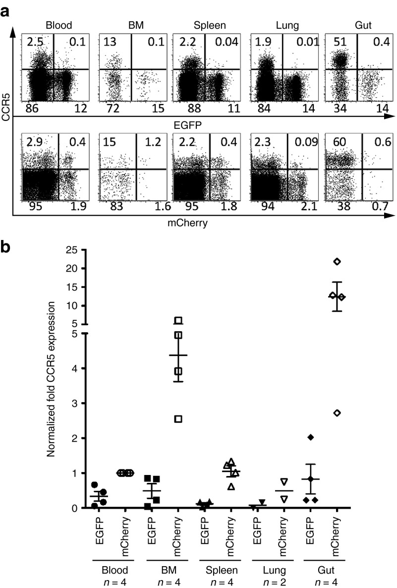
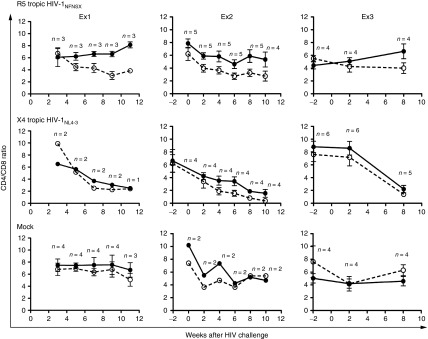
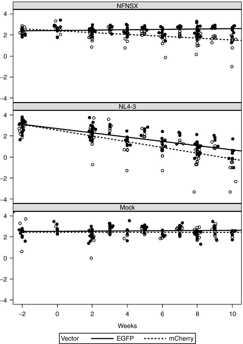
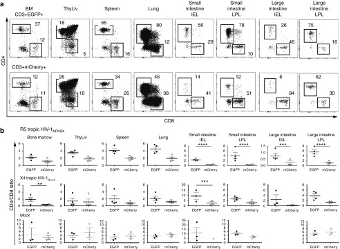
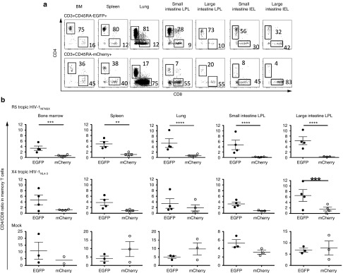
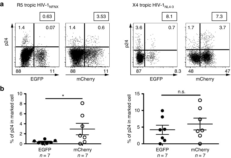
Transplantation of hematopoietic stem/progenitor cells (HSPC) modified with a lentiviral vector bearing a potent nontoxic short hairpin RNA (sh1005) directed to the HIV coreceptor CCR5 is capable of continuously producing CCR5 downregulated CD4+ T lymphocytes. Here, we characterized HIV-1 resistance of the sh1005-modified CD4+ T lymphocytes in vivo in humanized bone marrow/liver/thymus (hu BLT) mice. The sh1005-modified CD4+ T lymphocytes were positively selected in CCR5-tropic HIV-1-challenged mice. The sh1005-modified memory CD4+ T lymphocytes (the primary target of CCR5-tropic HIV-1) expressing sh1005 were maintained in lymphoid tissues in CCR5-tropic HIV-1-challenged mice. Frequencies of HIV-1 p24 expressing cells were significantly reduced in the sh1005-modified splenocytes by ex vivo cell stimulation confirming that CCR5 downregulated sh1005 modified cells are protected from viral infection. These results demonstrate that stable CCR5 downregulation through genetic modification of human HSPC by lentivirally delivered sh1005 is highly effective in providing HIV-1 resistance. Our results provide in vivo evidence in a relevant small animal model that sh1005 is a potent early-step anti-HIV reagent that has potential as a novel anti-HIV-1 HSPC gene therapeutic reagent for human applications.


Figures

Figure 1
Figure 1
CCR5 downregulation in EGFP+ CD4+ T cells in the reconstituted hu BLT mice. (a) Representative data for CCR5 expression in CD4+ T cells. Upper panel shows EGFP+ sh1005 vector marked CD4+ T cells and lower panel shows mCherry+ control vector marked CD4+ T cells. Numbers in each quadrant indicate the percentage. (b) The level of CCR5 expression was compared in EGFP+ and mCherry+ human CD4+ T cells in lymphoid tissues from multiple transplanted hu BLT mice. We normalized CCR5 expression level using the mean CCR5 expression in mCherry+ cells from peripheral blood (Blood) as 1. Samples were obtained between 21 and 26 weeks after human Thy/Liv implantation. Bar represents mean value; n indicates number of mice.
Figure 2
Figure 2
Selective advantage of sh1005-modified CD4+ T cells in peripheral blood following R5 tropic HIV-1 challenge. Hu BLT mice were challenged with R5-tropic HIV-1NFNSX (top panel), X4-tropic HIV-1NL4-3 (middle panel) or mock (bottom panel) in 3 independent cohort of experiments (Ex1, EX2 and EX3). Relative change in CD4/CD8 ratio kinetics in EGFP+ (solid circle and solid line) and in mCherry+ (open circle and dashed line) human CD3+ T lymphocytes in peripheral blood were compared between −2 and 11 weeks after HIV-1 challenge. Error bar shows standard deviation. n, number of mice.
Figure 3
Figure 3
Model slopes of CD4/CD8 ratio in peripheral blood. The relative changes in CD4/CD8 ratio of sh1005- (EGFP+) and no-shRNA control- (mCherry+) modified CD4+ T lymphocytes in peripheral blood were compared using a linear mixed effects model by including all data from the three cohort experiments (Ex1, EX2 and EX3). CD4/CD8 ratio in shRNA-transduced cells (EGFP+, dotted line) and control cells (mCherry, solid line) overlayed on raw values (EGFP+, open circle; mCherry+, solid circle) were shown.
Figure 4
Figure 4
Maintenance of CD4/CD8 ratio in sh1005-modified CD4+ T cells in lymphoid tissue. (a) Representative data for percentage of CD4 and CD8 expression in EGFP+ (top panels) and mCherry+ (bottom panels) CD3+ T lymphocytes in bone marrow (BM), spleen, lung, small and large intestine (SI and LI) and intraepithelial lymphocytes (IEL) and lamina propria lymphocytes (LPL) in a R5 tropic HIV-1NFNSX–challenged hu BLT mice. CD4+ and CD8+ populations were gated and percentages are shown. (b) The data of CD4/CD8 ratio in EGFP+ CD3+ T lymphocytes (solid circle) and in mCherry+ lymphocytes (open circle) from all analyzed mice are shown. Samples were obtained between 8 and 12 weeks after HIV-1 challenge. A bar represents the mean value. **P ≤ 0.01. ***P ≤ 0.001. ****P ≤ 0.0001.
Figure 5
Figure 5
Protection of CCR5 downregulated CD4+ memory T lymphocytes from R5-tropic HIV-1. (a) Percentage of central memory (CD45RA-/CD27+) in EGFP+ (Top panel) or mCherry+ (Bottom panel) CD4+ T lymphocytes in bone marrow (BM), spleen, lung, small and large intestine (SI and LI) lamina propria lymphocytes (LPL) and intraepithelial lymphocytes (IEL) from a R5 tropic HIV-1 infected hu BLT mouse. TCM indicates central memory T cells; TEM, effector memory T cells; and TTD, terminally differentiated cells. (b) Comparison of the level of % memory CD4+ T lymphocytes in EGFP+ (solid circle) and mCherry+ (open circle) from all analyzed mice (HIV-1NFNSX infected mice; n = 4, HIV-1NL4-3 infected mice; n = 4, mock infected mice; n = 3). A bar represents the mean value. ***P ≤ 0.001. ****P ≤ 0.0001.
Figure 6
Figure 6
Maintenance of CD4/CD8 ratio in sh1005 modified memory T cells in lymphoid tissues. (a) Representative data showing percentage of CD4 and CD8 expression in EGFP+ (top panels) and mCherry+ (bottom panels) memory T lymphocytes in bone marrow (BM), spleen, lung, small and large intestine (SI and LI) lamina propria lymphocytes (LPL) and intraepithelial lymphocytes (IEL) in a R5 tropic HIV-1NFNSX–challenged hu BLT mice. CD4+ and CD8+ populations were gated and percentages are shown. (b) CD4/CD8 ratios in EGFP+ memory T lymphocytes (solid circle) and in mCherry+ lymphocytes (open circle) from all analyzed mice are shown. A bar represents the mean value. **P ≤ 0.01. ***P ≤ 0.001. ****P ≤ 0.0001.
Figure 7
Figure 7
Protection of sh1005-modified splenocytes. Comparison of p24 reactivation in sh1005 (EGFP+) and no shRNA (mCherry+) population ex vivo. Splenocytes were isolated from R5-tropic HIV-1NFNSX–challenged hu BLT mice (n = 7) or X4-tropic HIV-1NL4-3–challenged hu BLT mice (n = 7). Splenocytes were isolated, stimulated with PHA/IL-2 for 5 days to reactivate HIV-1 and stained with anti-HIV-1 p24 gag monoclonal antibodies. (a) Representative p24 intracellular staining data in CD45+CD3+CD8- cell population were shown. (b) Results of p24 reactivation in EGFP+ population (solid circle) and in mCherry+ population (open circle) from all analyzed mice are shown. The percentages of p24 expression within EGFP+ or mCherry+ populations were normalized based on % each marker (EGFP or mCherry) expression for the comparison. *P ≤ 0.05. n.s., not significant.

References

    1. Kiem HP, Jerome KR, Deeks SG, McCune JM. Hematopoietic-stem-cell-based gene therapy for HIV disease. Cell Stem Cell. 2012;10:137–147. - PMC - PubMed
    1. Kitchen SG, Shimizu S, An DS. Stem cell-based anti-HIV gene therapy. Virology. 2011;411:260–272. - PMC - PubMed
    1. Hoxie JA, June CH.2012Novel cell and gene therapies for HIV Cold Spring Harb Perspect Med 2(10)doi:10.1101/cshperspect.a007179. - PMC - PubMed
    1. Baltimore D. Gene therapy. Intracellular immunization. Nature. 1988;335:395–396. - PubMed
    1. Anderson JS, Bauer G. Fighting HIV with stem cell therapy: one step closer to human trials. Expert Rev Anti Infect Ther. 2012;10:1071–1073. - PubMed