Review
. 2015 Mar 2;125(3):918-25.
doi: 10.1172/JCI76303. Epub 2015 Feb 17.

Emerging roles for enteric glia in gastrointestinal disorders

Review

Emerging roles for enteric glia in gastrointestinal disorders

Keith A Sharkey. J Clin Invest. .

Abstract

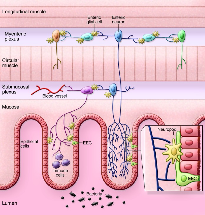
Enteric glia are important components of the enteric nervous system (ENS) and also form an extensive network in the mucosa of the gastrointestinal (GI) tract. Initially regarded as passive support cells, it is now clear that they are actively involved as cellular integrators in the control of motility and epithelial barrier function. Enteric glia form a cellular and molecular bridge between enteric nerves, enteroendocrine cells, immune cells, and epithelial cells, depending on their location. This Review highlights the role of enteric glia in GI motility disorders and in barrier and defense functions of the gut, notably in states of inflammation. It also discusses the involvement of enteric glia in neurological diseases that involve the GI tract.
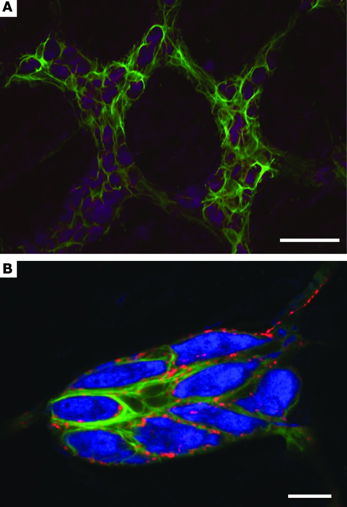

Figures

Figure 2
Figure 2. Enteric glia in the myenteric and submucosal plexus.
(A) Enteric glia labeled with GFAP (green) surround enteric neurons labeled with Hu C/D (purple) in the myenteric plexus of the mouse colon. Note that the expression of GFAP is not uniform in the myenteric plexus. Scale bar: 50 μm. (B) Enteric glia labeled with GFAP (green) surround enteric neurons labeled with Hu C/D (blue) in the submucosal plexus of the rat colon. Enteric glia and neurons are decorated with punctate nerve terminals expressing calcitonin gene–related peptide (red). Scale bar: 20 μm.
Figure 1
Figure 1. Schematic representation of the distribution of enteric glia in the GI tract.
Subpopulations of enteric glia are located around all classes of neurons in the myenteric and submucosal plexuses and in the mucosa. In the mucosa, enteric glia lie below the epithelium and connect to the “neuropod” of enteroendocrine cells (EECs) to form a tripartite connection among enteric nerves, enteroendocrine cells, and enteric glia (see text for details). Enteric glia also form a functional bridge between immune cells. Dark blue indicates intrinsic primary afferent neurons; light blue, interneurons; orange, excitatory motor neurons; green, inhibitory motor neurons; purple, secretomotor neurons.

References

    1. Kurahashi M, et al. A functional role for the ‘fibroblast-like cells’ in gastrointestinal smooth muscles. J Physiol. 2011;589(pt 3):697–710. - PMC - PubMed
    1. Muller PA, et al. Crosstalk between muscularis macrophages and enteric neurons regulates gastrointestinal motility. Cell. 2014;158(2):300–313. doi: 10.1016/j.cell.2014.04.050. - DOI - PMC - PubMed
    1. Furness JB. The enteric nervous system and neurogastroenterology. Nat Rev Gastroenterol Hepatol. 2012;9(5):286–294. doi: 10.1038/nrgastro.2012.32. - DOI - PubMed
    1. Gulbransen BD, Sharkey KA. Novel functional roles for enteric glia in the gastrointestinal tract. Nat Rev Gastroenterol Hepatol. 2012;9(11):625–632. doi: 10.1038/nrgastro.2012.138. - DOI - PubMed
    1. Gershon MD, Rothman TP. Enteric glia. Glia. 1991;4(2):195–204. doi: 10.1002/glia.440040211. - DOI - PubMed

Publication types

MeSH terms