. 2015 Feb 17;10(2):e0116515.
doi: 10.1371/journal.pone.0116515. eCollection 2015.

The acetate/ACSS2 switch regulates HIF-2 stress signaling in the tumor cell microenvironment

Affiliations

The acetate/ACSS2 switch regulates HIF-2 stress signaling in the tumor cell microenvironment

Rui Chen et al. PLoS One. .

Erratum in

Abstract

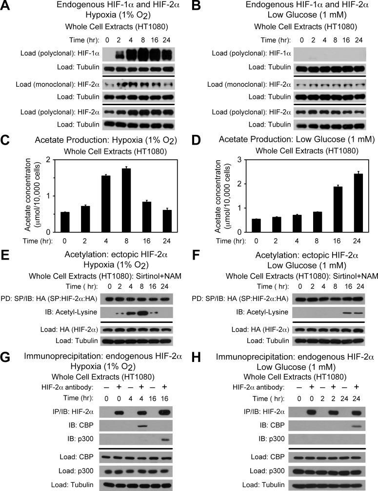
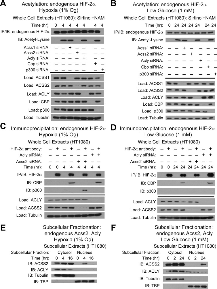
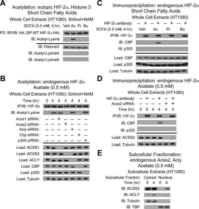
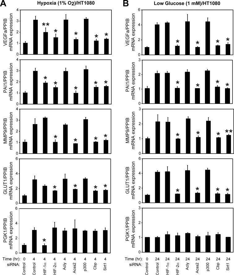
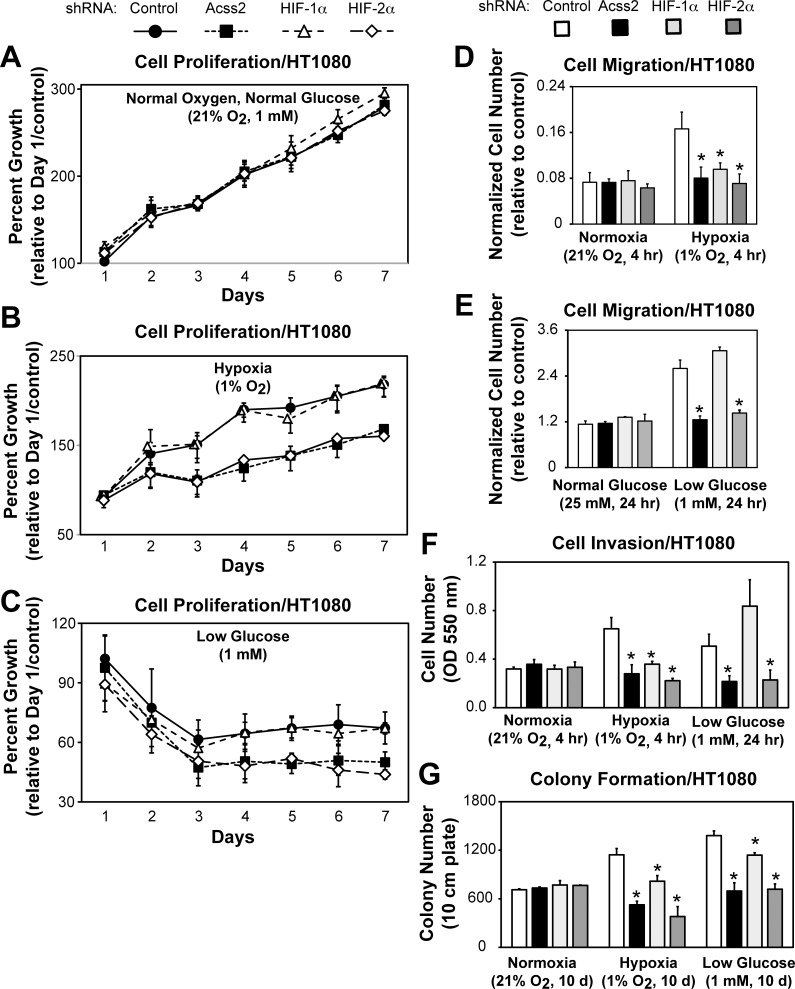
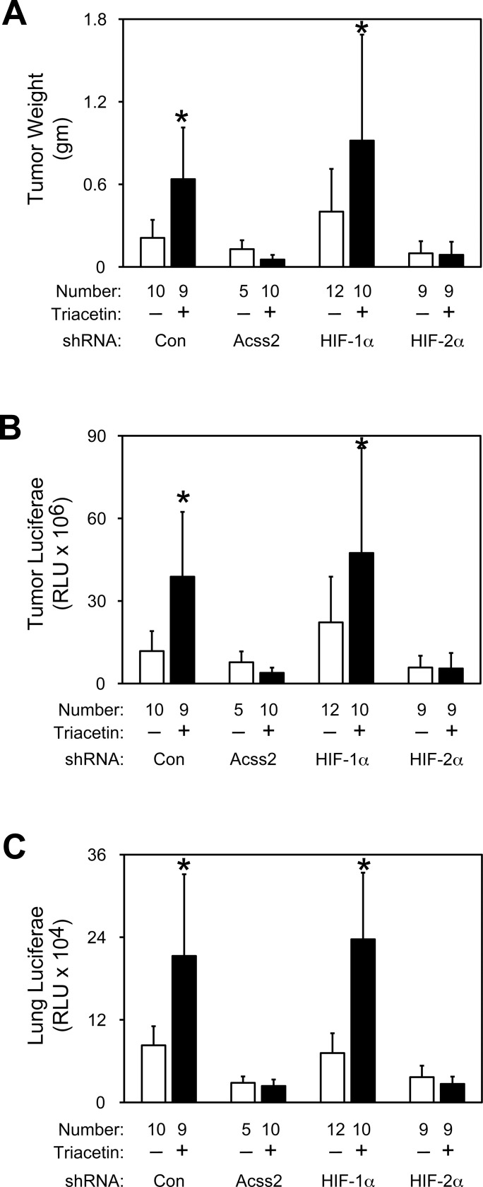
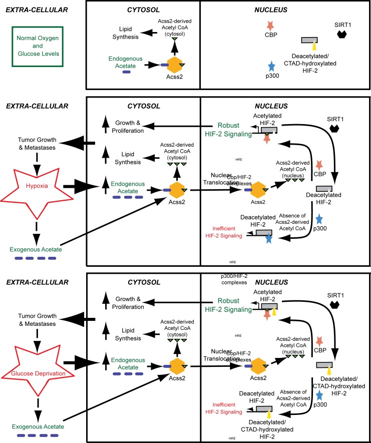
Optimal stress signaling by Hypoxia Inducible Factor 2 (HIF-2) during low oxygen states or hypoxia requires coupled actions of a specific coactivator/lysine acetyltransferase, Creb binding protein (CBP), and a specific deacetylase, Sirtuin 1 (SIRT1). We recently reported that acetylation of HIF-2 by CBP also requires a specific acetyl CoA generator, acetate-dependent acetyl CoA synthetase 2 (ACSS2). In this study, we demonstrate that ACSS2/HIF-2 signaling is active not only during hypoxia, but also during glucose deprivation. Acetate levels increase during stress and coincide with maximal HIF-2α acetylation and CBP/HIF-2α complex formation. Exogenous acetate induces HIF-2α acetylation, CBP/HIF-2α complex formation, and HIF-2 signaling. ACSS2 and HIF-2 are required for maximal colony formation, proliferation, migration, and invasion during stress. Acetate also stimulates flank tumor growth and metastasis in mice in an ACSS2 and HIF-2 dependent manner. Thus, ACSS2/CBP/SIRT1/HIF-2 signaling links nutrient sensing and stress signaling with cancer growth and progression in mammals.


Conflict of interest statement

Competing Interests: The authors have declared that no competing interests exist.

Figures

Fig 1
Fig 1. Acetate controls CBP/HIF-2α interactions during stress.
(A) Detection of endogenous HIF-1α or HIF-2α in whole cell extracts by immunoblotting after the indicated period of hypoxia exposure. Alpha-tubulin levels for each immunoblot are also shown. (B) Same as (A) except after low glucose exposure. (C) Cellular acetate levels generated after the indicated period of hypoxia exposure (n = 3 biological replicates/time-point; single measurement/replicate; mean/SD). (D) Same as (C) except after low glucose exposure. (E) Acetylation of ectopic HA-tagged HIF-2α after pulldown (PD) and detection by immunoblotting (IB) after the indicated period of hypoxia exposure with pharmacological inhibition of Sirt1 by sirtinol and nicotinamide (NAM). Levels of ectopic HIF-2α and alpha-tubulin levels from whole cell extracts prepared under the same conditions are also shown. (F) Same as (E) except after low glucose exposure. (G) Detection of endogenous CBP/HIF-2α or p300/HIF-2α complexes by immunoblotting (IB) after early (4 hr) or late (16 hr) hypoxia exposure. (H) Same as (G) except after early (2 hr) or late (24 hr) low glucose exposure. All experiments performed with HT1080 whole cell extracts.
Fig 2
Fig 2. ACSS2 is the molecular mediator of the acetate switch.
(A) Acetylation of endogenous HIF-2α after immunoprecipitation (IP) and detection by immunoblotting (IB) after early (4 hr) hypoxia exposure with pharmacological inhibition of Sirt1 by sirtinol and nicotinamide (NAM). (B) Same as (A) except after late (24 hr) low glucose exposure. (C) Detection of endogenous CBP/HIF-2α or p300/HIF-2α complexes by immunoblotting (IB) after early (4 hr) hypoxia exposure. (D) Same as (C) except after late (24 hr) low glucose exposure. (E) Detection of ACSS2 or ACLY in HT1080 subcellular fractions by immunoblotting (IB) after early (4 hr) or late (16 hr) hypoxia exposure. (F) Same as (E) except after early (2 hr) or late (24 hr) low glucose exposure. Experiments in (A)-(D) performed with HT1080 whole cell extracts.
Fig 3
Fig 3. Acetate induces ACSS2-dependent CBP/HIF-2α interactions.
(A) Acetylation of ectopic HA-tagged wild-type HIF-2α after pulldown (PD) and detection by immunoblotting (IB) after (4 hr) incubation with vehicle (Veh), or the short chain fatty acids acetate (Ac), propionate (Pr), and butyrate (Bu) with pharmacological inhibition of Sirt1 by sirtinol and nicotinamide (NAM). (B) Acetylation of endogenous HIF-2α after immunoprecipitation (IP) and detection by immunoblotting (IB) after (4 hr) incubation with acetate and following control, ACSS1, ACSS2, ACLY, CBP or p300 knockdown. (C) Endogenous CBP/HIF-2α or p300/HIF-2α complexes after (4 hr) treatment with vehicle (Veh), acetate (Ac), propionate (Pr), or butyrate (Bu). (D) Endogenous CBP/HIF-2α or p300/HIF-2α complexes detected by immunoblotting (IB) after (4 hr) acetate exposure following ACSS2 knockdown. (E) Detection of ACSS2 or ACLY in HT1080 subcellular fractions by immunoblotting (IB) after (4 hr) acetate exposure. Experiments in (A)-(D) performed with HT1080 whole cell extracts.
Fig 4
Fig 4. ACSS2, CBP, and SIRT1 are required for HIF-2 signaling.
Semi-quantitative RTPCR measurement of HIF-1 selective (PGK1), HIF-2 selective (MMP9, GLUT1), or HIF-1/HIF-2 co-regulated (VEGFa, PAI1) target genes following HIF-1α, HIF-2α, ACLY, ACSS2, p300, CBP, or SIRT1 knockdown and after (A) early (4 hr) hypoxia exposure, or (B) after incubation under late (24 hr) low glucose conditions. Comparison by one-tailed t-test between control knockdown/treatment and specified knockdown/treatment with significant reductions compared to control indicated (single pool of triplicate biological replicates/manipulation; triplicate measurements/pool; mean/SD; *, P<0.05; **, P<0.10).
Fig 5
Fig 5. ACSS2/CBP mediate acetate augmentation of HIF-2 signaling.
HIF target genes induced in (A) HT1080 cells expressing no ectopic HIF (control), oxygen-insensitive (PPN) HIF-1α, or PPN HIF-2α following control or ACSS2 knockdown, and treated with vehicle or acetate, or in (B) HT1080 cells expressing PPN HIF-2α following control, p300, CBP, or ACSS2 knockdown, and treated with vehicle or acetate. Comparison by one-tailed t-test between vehicle (empty bars) and acetate treatment (filled bars) with significant increases compared to vehicle treatment indicated (single pool of triplicate biological replicates/manipulation; triplicate measurements/pool; mean/SD; *, P<0.05; **, P<0.10).
Fig 6
Fig 6. ACSS2 and HIF-2α regulate in vitro tumor cell properties.
Cell proliferation assessed by cell survival for stably transformed HT1080 cells exposed to either (A) ambient, (B) hypoxic, or (C) low glucose conditions (n = 8/treatment/day) and expressing control (black circles), ACSS2 (black squares), HIF-1α (white triangles), or HIF-2α (white diamonds) shRNA. Cell migration assessed for same control (white bars), ACSS2 (black bars), HIF-1α (light gray bars), or HIF-2α (dark gray bars) knockdown cells exposed to either (D) hypoxic or (E) low glucose conditions (n = 3/treatment). (F) Cell invasion assessed under ambient, hypoxic, or low glucose conditions (n = 3/treatment). (G) Colony formation assessed under ambient, hypoxic, or low glucose conditions (n = 3/treatment). Comparison by one-tailed t-test between control and specified knockdown/treatment with significant reductions compared to control indicated (mean/SD for indicated number of replicates; *, P<0.05).
Fig 7
Fig 7. ACSS2 and HIF-2α regulate in vivo tumor cell properties.
(A) Weights of flank tumors derived from HT1080 cells expressing control (white bars), ACSS2 (black bars), HIF-1α (light gray bars), or HIF-2α (dark gray bars) shRNA downstream of a luciferase cDNA cassette. Tumor-bearing mice received daily vehicle or acetate (triacetin) oral gavage treatments beginning 6 days after tumor cell implantation until euthanization (20 days following tumor cell implantation). (B) Luciferase activity of flank tumor or (C) lung extracts from same mice in (A). For (A) through (C), comparison by one-tailed t-test or z-test between vehicle and acetate treatments with significant increases compared to vehicle treatment indicated (mean/SD for indicated number of mice with tumors; *, P<0.05). Not indicated is the tumor failure mice (no tumor detected at the completion of the experiment), which were as follows: n = 2 control shRNA/vehicle, n = 7 ACSS2 shRNA/vehicle, n = 0 HIF-1α shRNA/vehicle, or n = 3 HIF-2α shRNA/vehicle, n = 0 control shRNA/triacetin, n = 3 ACSS2 shRNA/triacetin, n = 2 HIF-1α shRNA/triacetin, or n = 4 HIF-2α shRNA/triacetin.
Fig 8
Fig 8. Proposed role of the acetate switch in tumor biology.
Endogenous acetate generated in response to hypoxia or glucose deprivation, or exogenous acetate originating from neighboring cells or from gastrointestinal uptake, stimulates ACSS2-dependent acetyl CoA production in the cytosol, but also directs production of an acetyl CoA pool that is localized in the nucleus upon ACSS2 nuclear translocation. In the cytosol, ACSS2 contributes to lipid synthesis, likely for cell growth and proliferation. In the nucleus, the acetyltransferase/coactivator CBP uses this specific ACSS2-derived acetyl CoA pool for HIF-2α acetylation and CBP/HIF-2α complex formation, which augments HIF-2 signaling. CBP is only bound to HIF-2α while it is undergoing the acetylation reaction, which occurs as long as ACSS2-generated acetyl CoA remains available. When acetylation of HIF-2α is complete, CBP is released. SIRT1 then deacetylates HIF-2α, restoring HIF-2α to a CBP substrate. After transformation or as a tumor forms, acetate may be produced constitutively, which further augments tumor growth. In the absence of ACSS2-generated acetyl CoA, HIF-2α complexes with p300 during hypoxia, but not during glucose deprivation. The p300/HIF-2α complex, however, is inefficient at inducing HIF-2 signaling compared to CBP/HIF-2α.

Similar articles

Cited by

References

    1. Semenza GL (2009) Regulation of oxygen homeostasis by hypoxia-inducible factor 1. Physiology (Bethesda) 24: 97–106. 10.1152/physiol.00045.2008 - DOI - PubMed
    1. Chae KS, Kang MJ, Lee JH, Ryu BK, Lee MG, et al. (2011) Opposite functions of HIF-alpha isoforms in VEGF induction by TGF-beta1 under non-hypoxic conditions. Oncogene 30: 1213–1228. 10.1038/onc.2010.498 - DOI - PubMed
    1. Raval RR, Lau KW, Tran MG, Sowter HM, Mandriota SJ, et al. (2005) Contrasting properties of hypoxia-inducible factor 1 (HIF-1) and HIF-2 in von Hippel-Lindau-associated renal cell carcinoma. Mol Cell Biol 25: 5675–5686. - PMC - PubMed
    1. Imamura T, Kikuchi H, Herraiz MT, Park DY, Mizukami Y, et al. (2009) HIF-1alpha and HIF-2alpha have divergent roles in colon cancer. Int J Cancer 124: 763–771. 10.1002/ijc.24032 - DOI - PMC - PubMed
    1. Mahon PC, Hirota K, Semenza GL (2001) FIH-1: a novel protein that interacts with HIF-1alpha and VHL to mediate repression of HIF-1 transcriptional activity. Genes Dev 15: 2675–2686. - PMC - PubMed

Publication types