. 2015 Feb 17;5(2):e512.
doi: 10.1038/tp.2015.2.

A role for D-aspartate oxidase in schizophrenia and in schizophrenia-related symptoms induced by phencyclidine in mice

Affiliations

A role for D-aspartate oxidase in schizophrenia and in schizophrenia-related symptoms induced by phencyclidine in mice

F Errico et al. Transl Psychiatry. .

Abstract

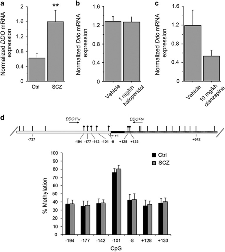
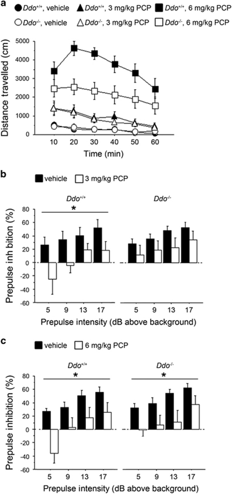
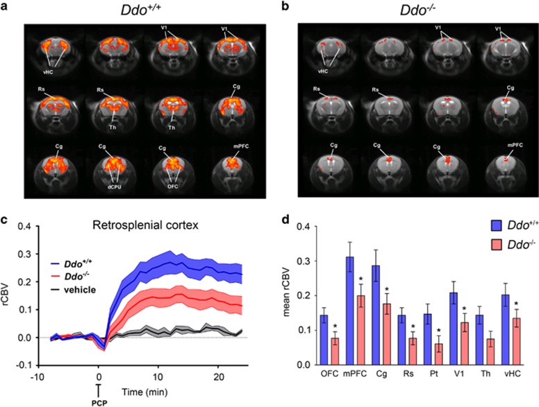
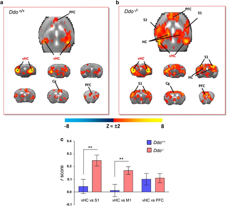
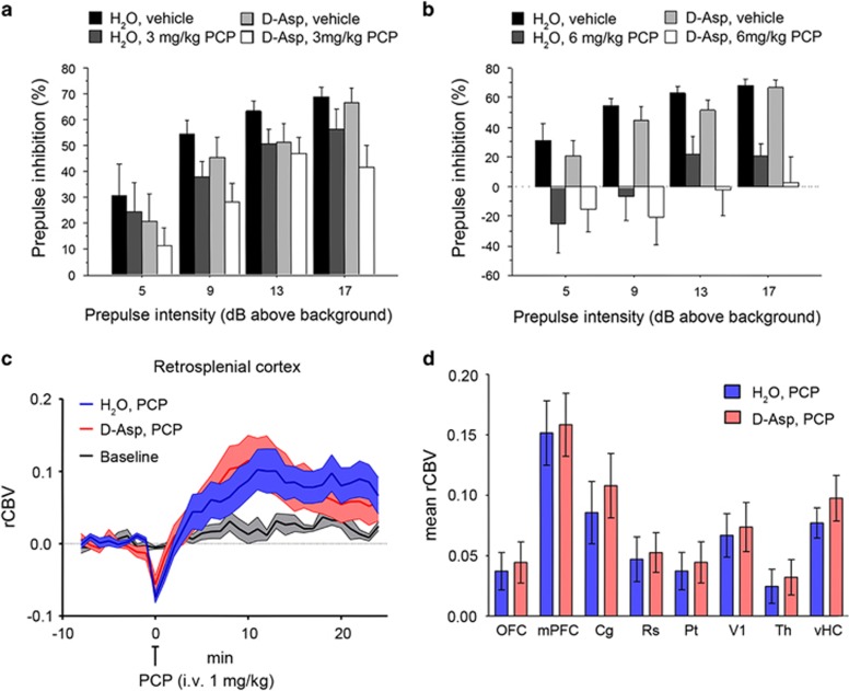
Increasing evidence points to a role for dysfunctional glutamate N-methyl-D-aspartate receptor (NMDAR) neurotransmission in schizophrenia. D-aspartate is an atypical amino acid that activates NMDARs through binding to the glutamate site on GluN2 subunits. D-aspartate is present in high amounts in the embryonic brain of mammals and rapidly decreases after birth, due to the activity of the enzyme D-aspartate oxidase (DDO). The agonistic activity exerted by D-aspartate on NMDARs and its neurodevelopmental occurrence make this D-amino acid a potential mediator for some of the NMDAR-related alterations observed in schizophrenia. Consistently, substantial reductions of D-aspartate and NMDA were recently observed in the postmortem prefrontal cortex of schizophrenic patients. Here we show that DDO mRNA expression is increased in prefrontal samples of schizophrenic patients, thus suggesting a plausible molecular event responsible for the D-aspartate imbalance previously described. To investigate whether altered D-aspartate levels can modulate schizophrenia-relevant circuits and behaviors, we also measured the psychotomimetic effects produced by the NMDAR antagonist, phencyclidine, in Ddo knockout mice (Ddo(-)(/-)), an animal model characterized by tonically increased D-aspartate levels since perinatal life. We show that Ddo(-/-) mice display a significant reduction in motor hyperactivity and prepulse inhibition deficit induced by phencyclidine, compared with controls. Furthermore, we reveal that increased levels of D-aspartate in Ddo(-/-) animals can significantly inhibit functional circuits activated by phencyclidine, and affect the development of cortico-hippocampal connectivity networks potentially involved in schizophrenia. Collectively, the present results suggest that altered D-aspartate levels can influence neurodevelopmental brain processes relevant to schizophrenia.


Figures

Figure 1
Figure 1
DDO mRNA expression and methylation of the DDO gene in the postmortem schizophrenia (SCZ) brain. (a) Analysis of DDO mRNA expression was performed by quantitative reverse transcription (qRT)-PCR in the PFC of SCZ patients (n=10) and control individuals (Ctrl, n=11). Quantity means of transcript were normalized to the geometric mean of three housekeeping genes. (b and c) Expression of Ddo mRNA, analyzed by qRT-PCR, in the PFC of mice on treatment with (b) 1 mg kg−1 haloperidol (n=6) or vehicle (n=5); (c) 10 mg kg−1 olanzapine (n=6) or vehicle (n=5). (d) DDO gene methylation analysis by NGS. The top panel shows the structure of the putative promoter of human DDO gene. The transcriptional start site (+1) is indicated by an arrow. The putative regulatory upstream region (white box), exon (black) and first intron (striped box) are indicated. The primer positions used for methylation analysis are indicated by arrows (DDO Fw and DDO Rv). Vertical bars represent the relative positions of each CpG site. Black circles represent the CpG sites analyzed (CpG −194, −177, −142, −101, −8, +128 and +133). The bar graph below shows the methylation degree of single CpG sites at DDO promoter in the PFC of 14 SCZ patients (gray bars) and 14 Ctrl subjects (black bars) at each CpG site. **P<0.01, ANCOVA. All the values are expressed as mean±s.e.m. DDO, D-aspartate oxidase; PFC, prefrontal cortex.
Figure 2
Figure 2
PCP-induced behavioral responses in Ddo−/− mice. (a) Motor activity induced by 3 mg kg−1 or 6 mg kg−1 PCP in Ddo+/+ (n=13 per treatment) and Ddo−/− mice (n=13 per treatment). Locomotion was expressed as distance traveled, measured in cm every 10 min over a 60-min session and presented as time course. (bc) PPI deficits induced by (b) 3 mg kg−1 or (c) 6 mg kg−1 PCP in Ddo+/+(n=10 per treatment) and Ddo−/− mice (n=10 per treatment). Percentage of the PPI was used as dependent variable and measured at different prepulse intensities (dB above 65 dB background level). *P<0.05, compared with vehicle control groups. All the values are expressed as the mean±s.e.m. DDO, D-aspartate oxidase; PCP, phencyclidine; PPI, prepulse inhibition.
Figure 3
Figure 3
PCP-induced fMRI response in Ddo−/− mice. (a and c) In Ddo+/+ mice, PCP elicited robust and sustained cortico–limbo–thalamic fMRI activation. (bd) This effect was strongly attenuated in Ddo−/− mice. Red/yellow in a and b indicates significant fMRI (rCBV) response to PCP (1 mg kg−1, intra-artery) with respect to vehicle (saline; 3.1<z-score<6, cluster correction threshold pc=0.001). *P<0.05, Student's t-test. Cg, cingulate cortex; dCPU, dorsal caudate putamen; DDO, D-aspartate oxidase; fMRI, functional magnetic resonance imaging; mPFC, medial prefrontal cortex; OFC, orbitofrontal cortex; PCP, phencyclidine; rCBV, relative cerebral blood volume; Rs, retrosplenial cortex; Th, thalamus; vHc, ventral hippocampus; V1, visual cortex.
Figure 4
Figure 4
Cortico–hippocampal connectivity in Ddo−/− mice. Transverse and coronal brain section heat maps showing voxels for which the fMRI BOLD signal was significantly correlated with the ventral hippocampus (seed regions in black) in (a) Ddo+/+and (b) Ddo−/− mice. The z-score indicates the strength of the correlation. (c) A stronger and more widespread profile of cortical (somatosensory) hippocampal connectivity was apparent in Ddo−/− mice. Strength (r-score) of cortico–hippocampal connectivity in representative regions of interest of Ddo+/+ and Ddo−/− mice. **P<0.01, Student's t-test. Cg, cingulate cortex; DDO, D-aspartate oxidase; fMRI, functional magnetic resonance imaging; PFC, prefrontal cortex; S1, somatosensory (parietal) cortex; vHc, ventral hippocampus; V1, visual cortex.
Figure 5
Figure 5
Effect of D-aspartate (D-Asp) supplementation on PCP-induced responses. (a and b) Prepulse inhibition responses to PCP after 1-month oral administration of D-aspartate in mice treated with (a) 3 mg kg−1 PCP (n=10 H2O; n=9 D-Asp) or vehicle (n=10 H2O; n=8 D-Asp), (b) 6 mg kg−1 PCP (n=10 H2O; n=10 D-Asp) or vehicle (n=10 H2O; n=8 D-Asp). Percentage of the PPI was used as dependent variable and measured at different prepulse intensities (shown as dB above 65 dB background level). All the values are expressed as the mean±s.e.m. (c and d) PCP-induced fMRI response in D-Asp- and H2O-treated adult C57BL6/J mice. In both groups of animals, PCP elicited robust and sustained cortico–limbo–thalamic fMRI activation (left and right panels). No statistically significant difference in the inter-group response to PCP was observed either at the voxel level (z>1.6, cluster corrected at P<0.01) or when integrated at the level of volumes of interest (right panel; P>0.27, all regions, Student's t-test). Cg, cingulate cortex; dCPU, dorsal caudate putamen; fMRI, functional magnetic resonance imaging; mPFC, medial prefrontal cortex; OFC, orbitofrontal cortex; PCP, phencyclidine; PPI, prepulse inhibition; rCBV, relative cerebral blood volume; Rs, retrosplenial cortex; Th, thalamus; vHc, ventral hippocampus; V1, visual cortex.

References

    1. Moghaddam B, Javitt D. From revolution to evolution: the glutamate hypothesis of schizophrenia and its implication for treatment. Neuropsychopharmacology. 2012;37:4–15. - PMC - PubMed
    1. Coyle JT. NMDA receptor and schizophrenia: a brief history. Schizophr Bull. 2012;38:920–926. - PMC - PubMed
    1. Coyle JT, Tsai G. NMDA receptor function, neuroplasticity, and the pathophysiology of schizophrenia. Int Rev Neurobiol. 2004;59:491–515. - PubMed
    1. Sawa A, Snyder SH. Schizophrenia: neural mechanisms for novel therapies. Mol Med. 2003;9:3–9. - PMC - PubMed
    1. Kristiansen LV, Huerta I, Beneyto M, Meador-Woodruff JH. NMDA receptors and schizophrenia. Curr Opin Pharmacol. 2007;7:48–55. - PubMed

Publication types

MeSH terms

LinkOut - more resources