. 2015 May;36(5):524-534.
doi: 10.1002/humu.22770. Epub 2015 Apr 6.

On human disease-causing amino acid variants: statistical study of sequence and structural patterns

Affiliations

On human disease-causing amino acid variants: statistical study of sequence and structural patterns

Marharyta Petukh et al. Hum Mutat. 2015 May.

Abstract

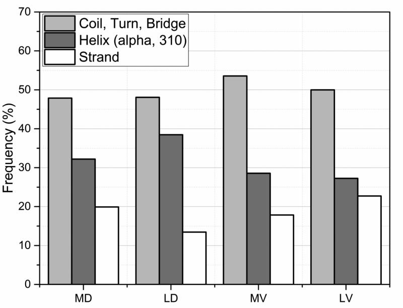
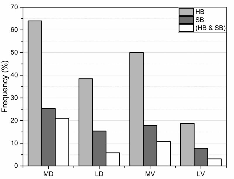
Statistical analysis was carried out on large set of naturally occurring human amino acid variations, and it was demonstrated that there is a preference for some amino acid substitutions to be associated with diseases. At an amino acid sequence level, it was shown that the disease-causing variants frequently involve drastic changes in amino acid physicochemical properties of proteins such as charge, hydrophobicity, and geometry. Structural analysis of variants involved in diseases and being frequently observed in human population showed similar trends: disease-causing variants tend to cause more changes in hydrogen bond network and salt bridges as compared with harmless amino acid mutations. Analysis of thermodynamics data reported in the literature, both experimental and computational, indicated that disease-causing variants tend to destabilize proteins and their interactions, which prompted us to investigate the effects of amino acid mutations on large databases of experimentally measured energy changes in unrelated proteins. Although the experimental datasets were linked neither to diseases nor exclusory to human proteins, the observed trends were the same: amino acid mutations tend to destabilize proteins and their interactions. Having in mind that structural and thermodynamics properties are interrelated, it is pointed out that any large change in any of them is anticipated to cause a disease.

Keywords: amino acid variations; disease mutations; hydrogen bond; structure.


Figures

Fig. 1
Fig. 1
Most frequent mutations. Frequency of top 26 mutations that make up 46% of HumVar database (disease and polymorphism). Rest of the 54% is made up by 246 mutations with 60 of them were observed only once.
Fig. 2
Fig. 2
Most frequent disease-causing mutations. Frequency of top 27 mutations that make up 53% of HumVar disease database. Rest of the 47% is composed of 245 mutations with 60 of them were observed only once.
Fig. 3
Fig. 3
The distribution of mutations sites within secondary structure elements (SSEs) for the four types of mutations sites MD, MV, LD and LV (see text for details).
Fig. 4
Fig. 4
Percentage of cases with changed (lost/gain of) hydrogen bonds (blue) and salt bridges (red) for the four types of mutations sites MD, MV, LD and LV (see text for details). The vertical axis indicates percentage of the cases the effect was found.
Fig. 5
Fig. 5
Degree of burial of WT residues classified three categories: exposed, partially exposed and buried. See text for more details for the burial classification and for MD, MV, LD and LV.
Fig. 6
Fig. 6
The mean of the experimental folding and binding free energy change caused by mutations taken from ProTherm and Skempi databases and grouped by physico-chemical property as: P<>P – polar to polar side chain substitution; C<>C – charged to charged; H<>H – hydrophobic to hydrophobic, C<>P – charged to polar and polar to charged; C<>H – charged to hydrophobic and hydrophobic to charged; P<>H – polar to hydrophobic and hydrophobic to polar. Charged residues (C): R, K, D and E; polar residues (P): S, T, N, and Q; hydrophobic residues (H): A, V, I, and L. Standard error is provided for each bar.
Fig. 7
Fig. 7
The median of experimental folding and binding free energy absolute change caused by mutations taken from ProTherm and Skempi databases and grouped according to largest effect and the rest of cases. Single arrow represents substitution for one type residue to another, as for example H>C is mutation from wild type hydrophobic to charged amino acid in the mutant. Charged residues (C): R, K, D, and E; polar residues (P): S, T, N, and Q; hydrophobic residues (H): A, V, I, and L. R and K are positively charged residues noted as C(+); while D and E – negatively charged residues noted as C(-). During C<>Ccons amino acid substitutions the charge of the residue conserves, that includes C(+)>C(+) and C(-)>C(-) groups of mutations.
Fig. 8
Fig. 8
Effect of the location of the mutated site on the change in binding/folding free energies. On the x-axis: the probability of the wild type residue being in the given location to cause large change in binding/folding free energy (perturbation index, Pp) due to mutation. On the y-axis: the averaged absolute value of experimentally obtained binding/folding free energies provided with standard error of mean as an error bar and the total number of cases across appropriate databases. Experimentally determined values of binding/folding free energies were obtained from extended Skempi and ProTherm databases respectively. Five location types of mutation sites were considered for binding energy change analysis (COR, RIM, SUP, SUR, and INT), and three – for folding free energy analysis (Buried, Partially Buried, and Exposed). For details see Method section.
Fig. 9
Fig. 9
Correlation between perturbation indexes (Pp) of the same mutation types causing change in binding and free energy (experimental values were obtained from extended Skempi and ProTherm databases respectively).

References

    1. Alexov E, Sternberg M. Understanding molecular effects of naturally occurring genetic differences. J Mol Biol. 2013;425(21):3911–3. - PubMed
    1. Atipairin A, Canyuk B, Ratanaphan A. Substitution of aspartic acid with glutamic acid at position 67 of the BRCA1 RING domain retains ubiquitin ligase activity and zinc (II) binding with a reduced transition temperature. JBIC Journal of Biological Inorganic Chemistry. 2011;16(2):217–226. - PubMed
    1. Bava KA, Gromiha MM, Uedaira H, Kitajima K, Sarai A. ProTherm, version 4.0: thermodynamic database for proteins and mutants. Nucleic Acids Res. 2004;32(Database issue):D120–1. - PMC - PubMed
    1. Baxa U, Steinbacher S, Weintraub A, Huber R, Seckler R. Mutations improving the folding of phage P22 tailspike protein affect its receptor binding activity. J Mol Biol. 1999;293(3):693–701. - PubMed
    1. Benedix A, Becker CM, de Groot BL, Caflisch A, Bockmann RA. Predicting free energy changes using structural ensembles. Nat Methods. 2009;6(1):3–4. - PubMed

Publication types

Substances

LinkOut - more resources