. 2015 May;83(5):1853-68.
doi: 10.1128/IAI.02833-14. Epub 2015 Feb 17.

Proteomic-based approach to gain insight into reprogramming of THP-1 cells exposed to Leishmania donovani over an early temporal window

Affiliations

Proteomic-based approach to gain insight into reprogramming of THP-1 cells exposed to Leishmania donovani over an early temporal window

Alok Kumar Singh et al. Infect Immun. 2015 May.

Abstract

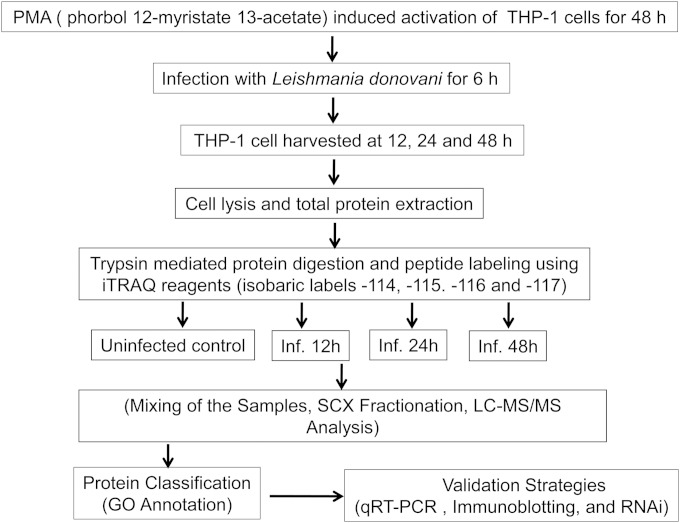
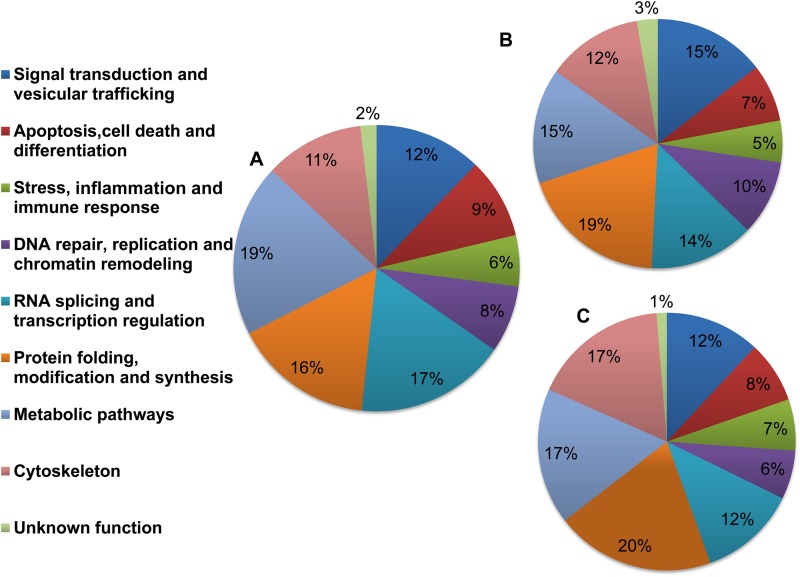
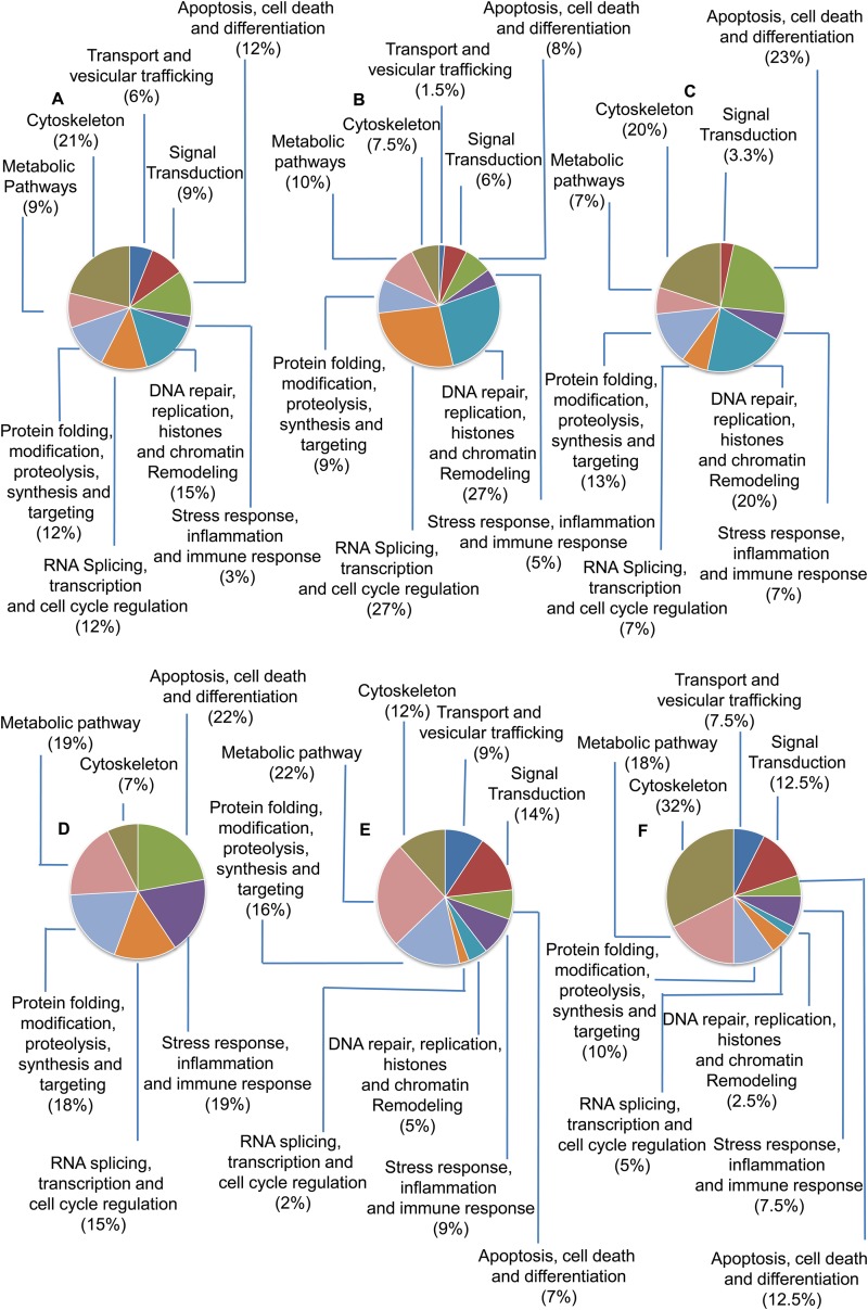
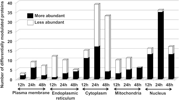
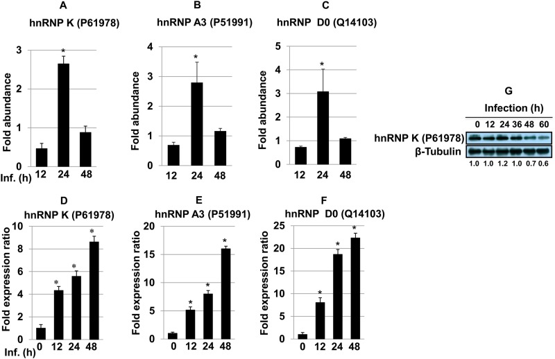
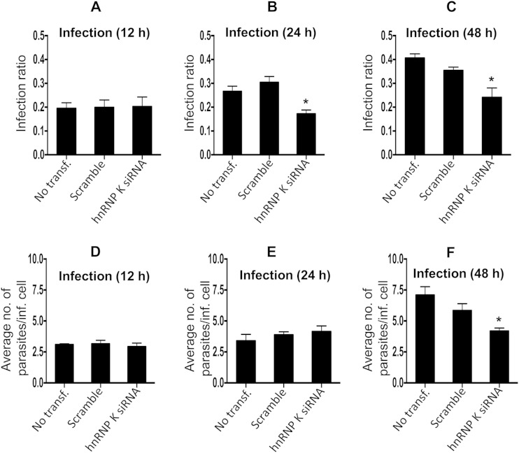
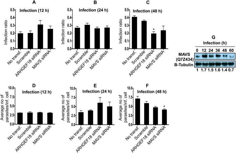
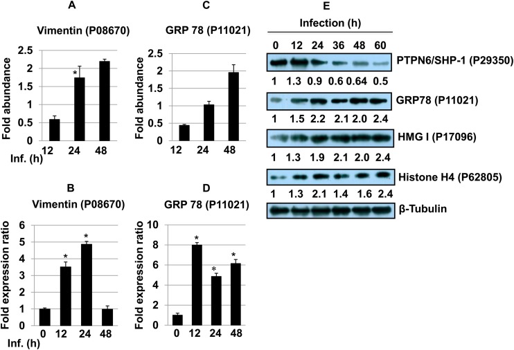
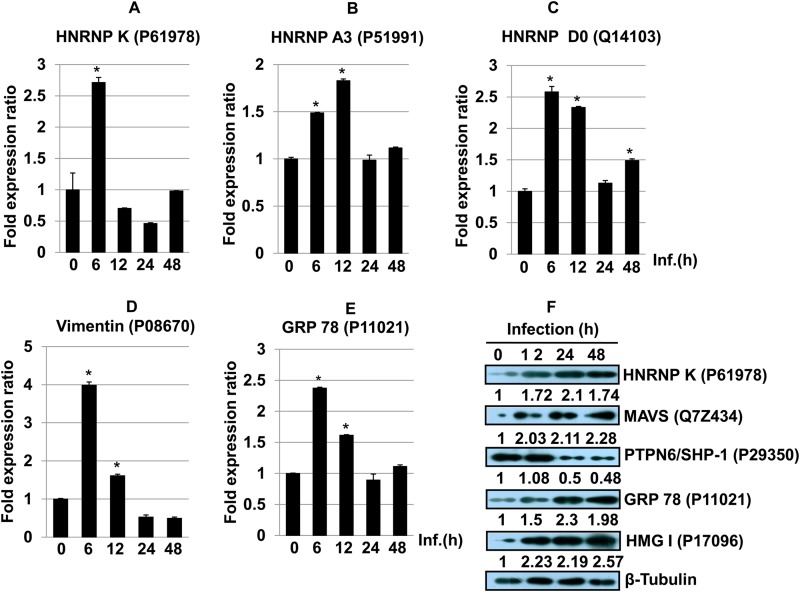
Leishmania donovani, a protozoan parasite, is the causative agent of visceral leishmaniasis. It lives and multiplies within the harsh environment of macrophages. In order to investigate how intracellular parasite manipulate the host cell environment, we undertook a quantitative proteomic study of human monocyte-derived macrophages (THP-1) following infection with L. donovani. We used the isobaric tags for relative and absolute quantification (iTRAQ) method and liquid chromatography-tandem mass spectrometry (LC-MS/MS) to compare expression profiles of noninfected and L. donovani-infected THP-1 cells. We detected modifications of protein expression in key metabolic pathways, including glycolysis and fatty acid oxidation, suggesting a global reprogramming of cell metabolism by the parasite. An increased abundance of proteins involved in gene transcription, RNA splicing (heterogeneous nuclear ribonucleoproteins [hnRNPs]), histones, and DNA repair and replication was observed at 24 h postinfection. Proteins involved in cell survival and signal transduction were more abundant at 24 h postinfection. Several of the differentially expressed proteins had not been previously implicated in response to the parasite, while the others support the previously identified proteins. Selected proteomics results were validated by real-time PCR and immunoblot analyses. Similar changes were observed in L. donovani-infected human monocyte-derived primary macrophages. The effect of RNA interference (RNAi)-mediated gene knockdown of proteins validated the relevance of the host quantitative proteomic screen. Our findings indicate that the host cell proteome is modulated after L. donovani infection, provide evidence for global reprogramming of cell metabolism, and demonstrate the complex relations between the host and parasite at the molecular level.


Figures

FIG 1
FIG 1
Workflow of the experimental strategy used in this study. Inf, infection.
FIG 2
FIG 2
Pie diagrams showing functional annotations and relative distributions of the total proteins detected in THP-1 cells infected with L. donovani. Nine major functional classes were identified based on their cellular function by using iTRAQ methodology as described in Materials and Methods. Percent distributions of the functionally annotated host proteins at 12 h (A), 24 h (B), and 48 h (C) after Leishmania infection are shown.
FIG 3
FIG 3
Functional distribution of differentially modulated host proteins (more and less abundant proteins) at 12, 24, and 48 h after Leishmania infection. (A to C) Percentages of more abundant proteins at 12 h (A), 24 h (B), and 48 h (C) postinfection. (D to F) Percentages of less abundant proteins at 12 h (D), 24 h (E), and 48 h (F) postinfection.
FIG 4
FIG 4
Gene Ontology-based classification of differentially abundant proteins present in different intracellular compartments.
FIG 5
FIG 5
(A to C) Bar diagrams showing differential abundances of hnRNP K (A), hnRNP A3 (B), and hnRNP D0 (C) in host macrophages after Leishmania infection at different time points. One-way ANOVA was applied to proteomics data to analyze the fold abundances. (D to F) The expression of hnRNP K (D), hnRNP A3 (E), and hnRNP D0 (F) was assessed by qRT-PCR. Real-time PCR data represent means from three independent experiments. Student's t test was applied to analyze the data. U6 small RNA (RNU6A) was used as an internal control. Data analysis was performed by using the 2−ΔΔCT method. *, P < 0.05. Inf., infection. (G) Western blot showing time-dependent expression of hnRNP K in THP-1 cells infected with L. donovani. Beta-tubulin was used as a loading control. Densitometric analysis shows the fold change in expression in infected THP-1 cells with respect to the untreated control group. The data are representative of three independent experiments. UniProt accession numbers are shown in parentheses.
FIG 6
FIG 6
(A to C) Bar diagrams showing the effect of gene knockdown of hnRNP K on the variation of the infection ratio at 12 h (A), 24 h (B), and 48 h (C). (D to F) Average number of parasites/infected cell at 12 h (D), 24 h (E), and 48 h (F). Results are representative of data from three separate experiments. Student's two-tailed t test was applied to analyze the data. *, P < 0.05. inf., infected; transf, transfection.
FIG 7
FIG 7
(A to C) Bar diagrams showing the effect of ARHGEF18 (Rho guanine nucleotide exchange factor 18) and MAVS (mitochondrial antiviral signaling protein) gene knockdown on the variation of the infection ratio at 12 h (A), 24 h (B), and 48 h (C). (D to F) Average number of parasites/infected cell at 12 h (D), 24 h (E), and 48 h (F). Results are representative of data from three separate experiments. Student's two-tailed t test was applied to analyze the data. *, P < 0.05. inf., infected; transf, transfection. (G) Western blot showing time-dependent expression of MAVS in THP-1 cells infected with L. donovani. Densitometric analysis shows the fold change in expression in infected THP-1 cells with respect to the untreated control group. The data are representative of three independent experiments.
FIG 8
FIG 8
(A and C) Bar diagrams showing differential abundances of vimentin (A) and the 78-kDa glucose-regulated protein (C) at different time points post-Leishmania infection. (B and D) The expression of vimentin (B) and 78-kDa glucose-regulated protein (D) was assessed by qRT-PCR. One-way ANOVA was applied to proteomics data to analyze the fold abundance. Real-time PCR data represent means from three independent experiments. Student's t test was applied to analyze the data. RNU6A was used as an internal control. Data analysis was performed by using the 2−ΔΔCT method. *, P < 0.05. Inf., infection. (E) Western blot analysis of PTPN6/SHP-1, GRP78, HMG-I, and histone H4 proteins across different time points in THP-1 cells infected with L. donovani. Beta-tubulin was used as a loading control. Densitometric analysis shows fold changes in expression in infected THP-1 cells with respect to the untreated control group. The data are representative of three independent experiments.
FIG 9
FIG 9
(A to E) Real-time PCR analyses of hnRNP K (A), hnRNP A3 (B), hnRNP D0 (C), vimentin (D), and GRP78 (E) across different time points in human monocyte-derived macrophages infected with L. donovani. The data are representative of three different biological experimental replicates. Student's t test was applied to analyze the data. RNU6A was used as an internal control. Data analysis was performed by using the 2−ΔΔCT method. *, P < 0.05. (F) Western blot analysis of hnRNP K, MAVS, PTPN6/SHP-1, GRP78, and HMG-I. Beta-tubulin was used as a loading control. Densitometric analysis shows fold changes in expression in infected THP-1 cells with respect to the untreated control group. The data are representative of three independent experiments.

Similar articles

Cited by

References

    1. Herwaldt BL. 1999. Leishmaniasis. Lancet 354:1191–1199. - PubMed
    1. Bogdan C, Röllinghoff M. 1999. How do protozoan parasites survive inside macrophages? Parasitol Today 15:22–28. doi:10.1016/S0169-4758(98)01362-3. - DOI - PubMed
    1. Olivier M, Gregory DJ, Forget G. 2005. Subversion mechanisms by which Leishmania parasites can escape the host immune response: a signaling point of view. Clin Microbiol Rev 18:293–305. doi:10.1128/CMR.18.2.293-305.2005. - DOI - PMC - PubMed
    1. Proudfoot L, O'Donnell CA, Liew FY. 1995. Glycoinositolphospholipids of Leishmania major inhibit nitric oxide synthesis and reduce leishmanicidal activity in murine macrophages. Eur J Immunol 25:745–750. doi:10.1002/eji.1830250318. - DOI - PubMed
    1. Forget G, Siminovitch KA, Brochu S, Rivest S, Radzioch D, Olivier M. 2001. Role of host phosphotyrosine phosphatase SHP-1 in the development of murine leishmaniasis. Eur J Immunol 31:3185–3196. doi:10.1002/1521-4141(200111)31:11<3185::AID-IMMU3185>3.0.CO;2-J. - DOI - PubMed

Publication types

LinkOut - more resources