Comparative Study
. 2015 May;473(5):1635-43.
doi: 10.1007/s11999-015-4192-2.

Platelet-rich concentrates differentially release growth factors and induce cell migration in vitro

Affiliations
Comparative Study

Platelet-rich concentrates differentially release growth factors and induce cell migration in vitro

Michael O Schär et al. Clin Orthop Relat Res. 2015 May.

Abstract

Background: Platelet-rich concentrates are used as a source of growth factors to improve the healing process. The diverse preparation protocols and the gaps in knowledge of their biological properties complicate the interpretation of clinical results.

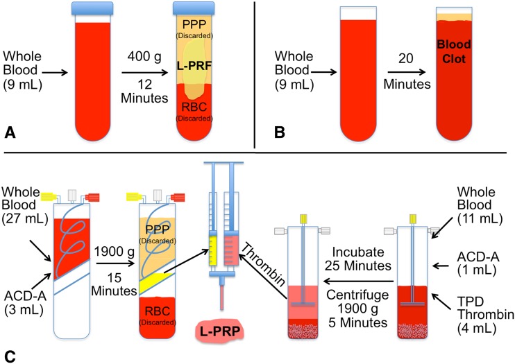
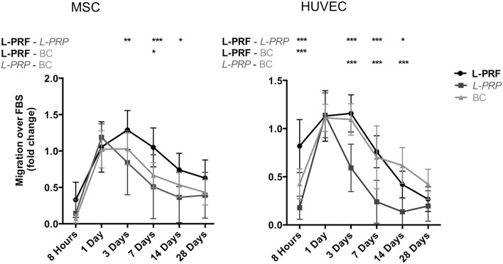
Questions/purposes: In this study we aimed to (1) analyze the concentration and kinetics of growth factors released from leukocyte- and platelet-rich fibrin (L-PRF), leukocyte- and platelet-rich plasma (L-PRP), and natural blood clot during in vitro culture; (2) investigate the migration of mesenchymal stem cells (MSCs) and human umbilical vein endothelial cells (HUVECs) as a functional response to the factors released; and (3) uncover correlations between individual growth factors with the initial platelet/leukocyte counts or the induced cell migration.

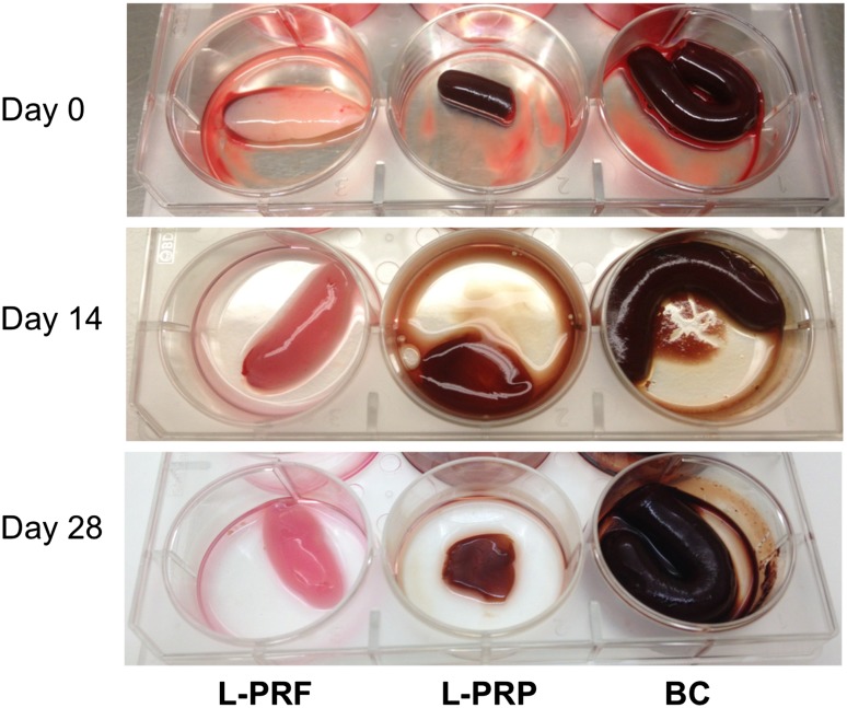
Methods: L-PRF, L-PRP, and natural blood clot prepared from 11 donors were cultured in vitro for 28 days and media supernatants collected after 8 hours and 1, 3, 7, 14, and 28 days. Released transforming growth factor β1 (TGF-β1), vascular endothelial growth factor (VEGF), insulin growth factor (IGF-1), platelet-derived growth factor AB (PDGF-AB), and interleukin-1β (IL-1β) were measured in the supernatants with enzyme-linked immunosorbent assay. Migration of MSC and HUVEC induced by the supernatants was evaluated in Boyden chambers.

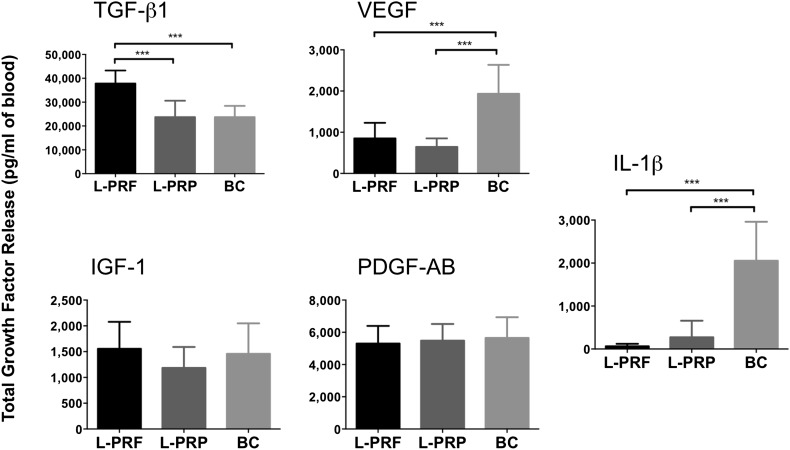
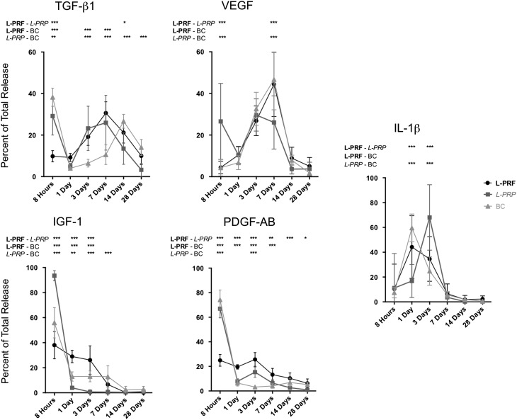
Results: More TGF-ß1 was released (mean ± SD in pg/mL of blood) from L-PRF (37,796 ± 5492) compared with L-PRP (23,738 ± 6848; p < 0.001) and blood clot (3739 ± 4690; p < 0.001), whereas more VEGF and IL-1ß were released from blood clot (1933 ± 704 and 2053 ± 908, respectively) compared with both L-PRP (642 ± 208; p < 0.001 and 273 ± 386; p < 0.001, respectively) and L-PRF (852 ± 376; p < 0.001 and 65 ± 56, p < 0.001, respectively). No differences were observed in IGF-1 and PDGF-AB released from any of the concentrates. TGF-β1 release peaked at Day 7 in L-PRF and at 8 hours and Day 7 in L-PRP and 8 hours and Day 14 in blood clot. In all concentrates, main release of VEGF occurred between 3 and 7 days and of IL-1β between Days 1 and 7. IGF-1 and PDGF-AB were released until Day 1 in L-PRP and blood clot, in contrast to sustained release over the first 3 days in L-PRF. The strongest migration of MSC occurred in response to L-PRF, and more HUVEC migration was seen in L-PRF and blood clot compared with L-PRP. TGF-β1 correlated with initial platelet counts in L-PRF (Pearson r = 0.66, p = 0.0273) and initial leukocyte counts in L-PRP (Pearson r = 0.83, p = 0.0016). A positive correlation of IL-1β on migration of MSC and HUVEC was revealed (Pearson r = 0.16, p = 0.0208; Pearson r = 0.31, p < 0.001).

Conclusions: In comparison to L-PRP, L-PRF had higher amounts of released TGF-β1, a long-term release of growth factors, and stronger induction of cell migration. Future preclinical studies should confirm these data in a defined injury model.

Clinical relevance: By characterizing the biologic properties of different platelet concentrates in vitro, we may gain a better understanding of their clinical effects and develop guidelines for specific future applications.


Figures

Fig. 1A–C
Fig. 1A–C
Schematic overview of the protocols to prepare platelet concentrates is shown. Each collected blood sample was divided and processed according to the depicted protocol to obtain (A) L-PRF; (B) blood clot, and (C) L-PRP concentrates.
Fig. 2
Fig. 2
The appearances of L-PRF, L-PRP, and blood clot (BC) during culture are shown. L-PRF, L-PRP, and blood clot concentrates were cultured in six-well plates for 28 days. The appearance of each concentrate at the time of preparation (Day 0), after Day 14, and Day 28 is shown.
Fig. 3
Fig. 3
Total growth factor release from L-PRF, L-PRP, and blood clot (BC) is shown. The total amount of TGF-ß1, VEGF, IGF-1, PDGF-AB, and IL-1ß released during the entire culturing period was determined by summing up the release of each growth factor measured in media cultured with L-PRF, L-PRP, or blood clot at each time point (8 hours and 1, 3, 7, 14, and 28 days). Data are presented as mean ± SD from triplicate measurements of 11 samples as total amount normalized to the volume of blood used for the preparation of the platelet concentrate. Statistical evaluation was done using one-way ANOVA and Bonferroni post hoc test. Significant differences are indicated: ***p < 0.001.
Fig. 4
Fig. 4
Kinetics of growth factor release from L-PRP, L-PRF, and blood clot (BC) are shown. Release of TGF-ß1, VEGF, IGF-1, PDGF-AB, and IL-1ß from cultured L-PRF, L-PRP, or BC was determined at each time point (8 hours, 1, 3, 7, 14 and 28 days). The amounts released are expressed as a percentage of total release per time point. Data are presented as mean ± SD from triplicate measurements of 11 samples. Statistical evaluation was done using two-way repeated ANOVA and Bonferroni post hoc test. Significant differences among the platelet concentrates for each factor at different time points are indicated: *p < 0.05, **p < 0.01, ***p < 0.001.
Fig. 5
Fig. 5
Migration of MSC and HUVEC in response to factors released from L-PRF, L-PRP, and blood clot (BC) is shown. Migration of MSC and HUVEC was assessed in Boyden chambers with media collected after 8 hours and 1, 3, 7, 14 and 28 days of cultured L-PRP, L-PRF, and blood clot compared with media containing 10% FBS and expressed as fold change. Data are presented as mean ± SD from triplicate measurements of 11 samples. Statistical evaluation was done using two-way repeated ANOVA and Bonferroni post hoc test. Significant differences for MSC and HUVEC migration among the platelet concentrates at different time points are indicated: *p < 0.05, **p < 0.01, ***p < 0.001.

References

    1. Ahmed TA, Dare EV, Hincke M. Fibrin: a versatile scaffold for tissue engineering applications. Tissue Eng Part B Rev. 2008;14:199–215. doi: 10.1089/ten.teb.2007.0435. - DOI - PubMed
    1. Bielecki T. Dohan Ehrenfest DM, Everts PA, Wiczkowski A. The role of leukocytes from L-PRP/L-PRF in wound healing and immune defense: new perspectives. Curr Pharm Biotechnol. 2012;13:1153–1162. doi: 10.2174/138920112800624373. - DOI - PubMed
    1. Bielecki TM, Gazdzik TS, Arendt J, Szczepanski T, Krol W, Wielkoszynski T. Antibacterial effect of autologous platelet gel enriched with growth factors and other active substances: an in vitro study. J Bone Joint Surg Br. 2007;89:417–420. doi: 10.1302/0301-620X.89B3.18491. - DOI - PubMed
    1. Boswell SG, Cole BJ, Sundman EA, Karas V, Fortier LA. Platelet-rich plasma: a milieu of bioactive factors. Arthroscopy. 2012;28:429–439. doi: 10.1016/j.arthro.2011.10.018. - DOI - PubMed
    1. Boswell SG, Schnabel LV, Mohammed HO, Sundman EA, Minas T, Fortier LA. Increasing platelet concentrations in leukocyte-reduced platelet-rich plasma decrease collagen gene synthesis in tendons. Am J Sports Med. 2014;42:42–49. doi: 10.1177/0363546513507566. - DOI - PubMed

Publication types

MeSH terms