. 2015 Aug;99(15):6315-26.
doi: 10.1007/s00253-015-6443-2. Epub 2015 Feb 18.

Discovery of novel S. aureus autolysins and molecular engineering to enhance bacteriolytic activity

Affiliations

Discovery of novel S. aureus autolysins and molecular engineering to enhance bacteriolytic activity

Daniel C Osipovitch et al. Appl Microbiol Biotechnol. 2015 Aug.

Abstract

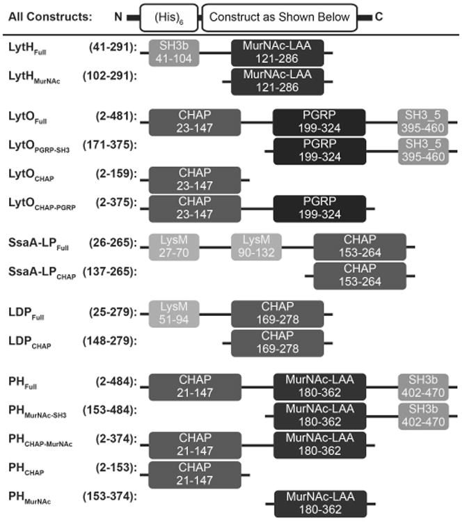
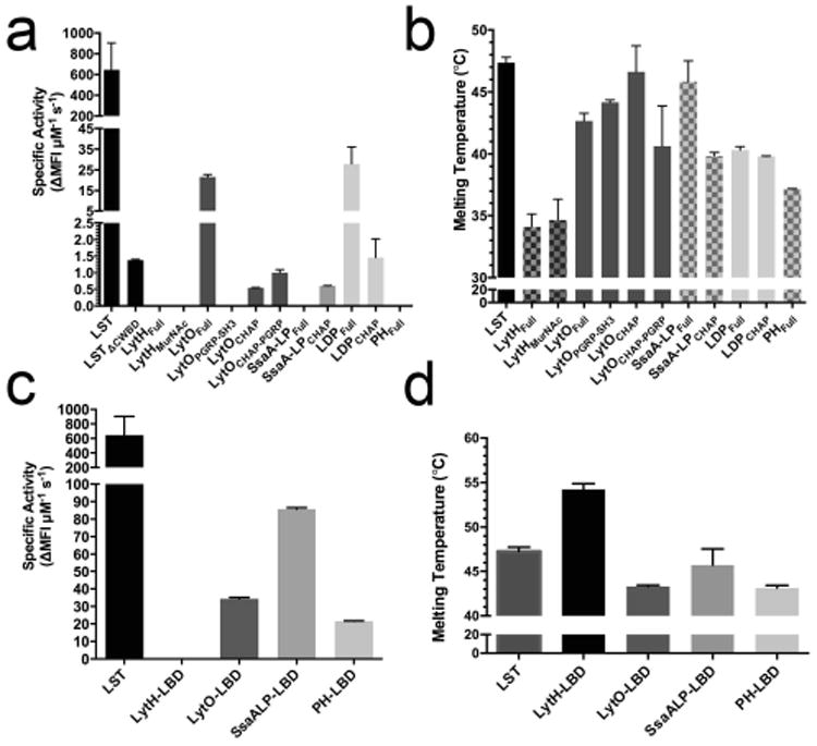
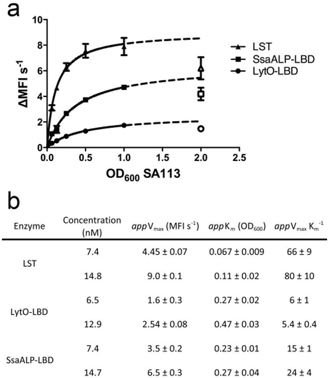
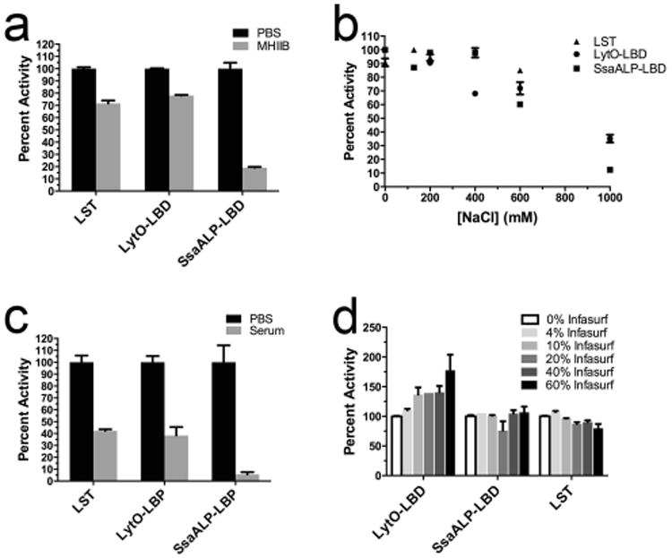
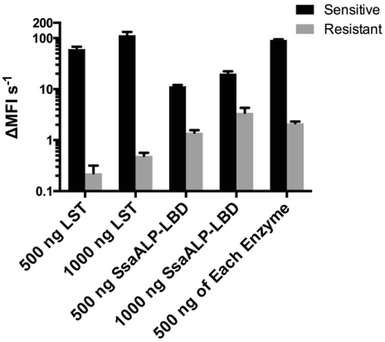
Staphylococcus aureus is a dangerous bacterial pathogen whose clinical impact has been amplified by the emergence and rapid spread of antibiotic resistance. In the search for more effective therapeutic strategies, great effort has been placed on the study and development of staphylolytic enzymes, which benefit from high potency activity toward drug-resistant strains, and a low inherent susceptibility to emergence of new resistance phenotypes. To date, the majority of therapeutic candidates have derived from either bacteriophage or environmental competitors of S. aureus. Little to no consideration has been given to cis-acting autolysins that represent key elements in the bacterium's endogenous cell wall maintenance and recycling machinery. In this study, five putative autolysins were cloned from the S. aureus genome, and their activities were evaluated. Four of these novel enzymes, or component domains thereof, demonstrated lytic activity toward live S. aureus cells, but their potencies were 10s to 1000s of times lower than that of the well-characterized therapeutic candidate lysostaphin. We hypothesized that their poor activities were due in part to suboptimal cell wall targeting associated with their native cell wall binding domains, and we sought to enhance their antibacterial potential via chimeragenesis with the peptidoglycan binding domain of lysostaphin. The most potent chimera exhibited a 140-fold increase in lytic rate, bringing it within 8-fold of lysostaphin. While this enzyme was sensitive to certain biologically relevant environmental factors and failed to exhibit a measurable minimal inhibitory concentration, it was able to kill lysostaphin-resistant S. aureus and ultimately proved active in lung surfactant. We conclude that the S. aureus proteome represents a rich and untapped reservoir of novel antibacterial enzymes, and we demonstrate enhanced bacteriolytic activity via improved cell wall targeting of autolysin catalytic domains.


Conflict of interest statement

The authors claim no conflict of interest.

Figures

Fig. 1
Fig. 1
Schematic of autolysin constructs. Numbers represent amino acid positions corresponding to the full length, wild type protein from UniProt. On the left is the construct name, followed by the corresponding amino acid numbers of the truncations. On the right is the illustration of each construct. Domain names and amino acid numbers were obtained from the NCBI Conserved Domain Search and abbreviations are as follows: CHAP, cysteine/histidine-dependent amidohydrolase/peptidase; MurNAc-LAA, N-acetylmuramoyl-L-alanine amidase; PGRP, peptidoglycan recognition proteins; SH3, bacterial SRC homology 3 domain; LysM, lysin motif. Dark greys represent purported catalytic domains and light greys represent purported cell wall binding domains. The function of the PGRP domain, black, is currently unknown.
Fig. 2
Fig. 2
Preliminary characterization of autolysins and chimeras. A) Specific activities of the autolysins against live SA113 cells as measured by the SYTOX fluorescence kinetic assay. Lysins were tested up to 50 μg per 250 μL reaction. The LSTΔcwbd designation represents the LST's catalytic domain, for which kinetic data was previously published (Osipovitch, 2014). B) Melting temperatures of autolysins using differential scanning fluorimetry. C) Specific activity of chimeric lysins against live SA113 cells as determined by the SYTOX fluorescent assay. The LBD designation represents LST binding domain chimeras. LytH-LBD was tested up to 100 μg (14 μM), and the concentrations of the other enzymes were as follows: LytO-LBD=800 ng (100 nM), SsaALP-LBD=700 ng (100 nM), and PH-LBD=1250 ng (162 nM). D) Melting temperatures of chimeras. ΔMFI s-1 (change in mean fluorescence intensity per second) represents the slope of the steepest linear region of the lysis curve. All error bars represent standard deviations from two or more technical replicates.
Fig. 3
Fig. 3
Pseudo-Michaelis-Menten kinetic analyses of LST and engineered autolysins. A) Curve fits used for determining pseudo-Michaelis-Menten constants. The dotted line represents an extrapolation of fit data, and the open shapes are values that were excluded in the curve fit. B) Table of apparent Michaelis-Menten parameters determined at two concentrations of each enzyme. Data are the means of triplicate measurements made in biological duplicate.
Fig. 4
Fig. 4
Specific activities across various concentrations of enzyme. A) LST B) LytO-LBD C) SsaALP-LBD. ΔMFI s-1 (change in mean fluorescence intensity per second) represents the slope of the steepest linear region of the lysis curve. Each data point represents the mean of duplicate measures (LST) or quadruplicate measures (LytO-LBD and SsaALP-LBD) from a representative experiment, and error bars are standard deviations. Biological replicates exhibited the same trends.
Fig. 5
Fig. 5
Environmental effects on the activity of LST and chimeric enzymes. Activities were measured with the SYTOX fluorescent kinetic assay and are reported as normalized percent activities. A) Activity in MHIIB medium supplemented with 2% NaCl. B) Activity in PBS supplemented with various salt concentrations. C) Activity in the presence of 60% human serum. D) Activity in the presence of the clinical-grade calf lung surfactant Infasurf. Error bars represent standard deviations, and all points include at least duplicate measurements.
Fig. 6
Fig. 6
SsaALP-LBD lytic activity towards LST sensitive and resistant strains. Rate of lysis (change in mean fluorescence intensity per second), measured by a fluorescence assay, is shown for the LST-sensitive strain RN4220/PLI50 and the LST-resistant strain RN4220/PLI50∷end epr (Gargis 2010, DeHart 1995). Note the log-scale axis. For both enzymes, 500 ng and 1000 ng correspond to 0.07 and 0.15 μM, respectively. Values represent background subtracted means from triplicate measurements run in biological duplicate, with error shown as standard deviation.

References

    1. Becker SC, Dong S, Baker JR, Foster-Frey J, Pritchard DG, Donovan DM. LysK CHAP endopeptidase domain is required for lysis of live staphylococcal cells. FEMS Microbiol Lett. 2009;294(1):52–60. doi: 10.1111/j.1574-6968.2009.01541.x. - DOI - PubMed
    1. Bose JL, Lehman MK, Fey PD, Bayles KW. Contribution of the Staphylococcus aureus Atl AM and GL murein hydrolase activities in cell division, autolysis, and biofilm formation. PLoS One. 2012;7(7):e42244. doi: 10.1371/journal.pone.0042244. - DOI - PMC - PubMed
    1. Brunskill EW, Bayles KW. Identification of LytSR-regulated genes from Staphylococcus aureus. J Bacteriol. 1996;178(19):5810–2. - PMC - PubMed
    1. Buist G, Steen A, Kok J, Kuipers OP. LysM, a widely distributed protein motif for binding to (peptido)glycans. Mol Microbiol. 2008;68(4):838–47. doi: 10.1111/j.1365-2958.2008.06211.x. - DOI - PubMed
    1. Centers for Disease Control and Prevention. Antibiotic Resistance Threats in the United States. 2013 Threats Report 2013

Publication types

MeSH terms

LinkOut - more resources