. 2015 Jun;99(11):4785-94.
doi: 10.1007/s00253-015-6445-0. Epub 2015 Feb 18.

The feasibility of using irreversible electroporation to introduce pores in bacterial cellulose scaffolds for tissue engineering

Affiliations

The feasibility of using irreversible electroporation to introduce pores in bacterial cellulose scaffolds for tissue engineering

Adwoa Baah-Dwomoh et al. Appl Microbiol Biotechnol. 2015 Jun.

Abstract

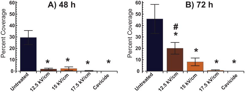
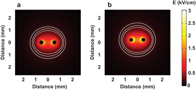
This work investigates the feasibility of the use of irreversible electroporation (IRE) in the biofabrication of 3D cellulose nanofibril networks via the bacterial strain Gluconacetobacter xylinus. IRE uses electrical pulses to increase membrane permeability by altering the transmembrane potential; past a threshold, damage to the cell becomes too great and leads to cell death. We hypothesized that using IRE to kill the bacteria at specific locations and particular times, we could introduce conduits in the overall scaffold by preventing cellulose biosynthesis locally. Through mathematical modeling and experimental techniques, electrical effects were investigated and the parameters for IRE of G. xylinus were determined. We found that for a specific set of parameters, an applied electric field of 8 to 12.5 kV/cm, producing a local field of 3 kV/cm, was sufficient to kill most of the bacteria and create a localized pore. However, an applied electric field of 17.5 kV/cm was required to kill all. Results suggest that IRE may be an effective tool to create scaffolds with appropriate porosity for orthopedic applications. Ideally, these engineered scaffolds could be used to successfully treat osteochondral defects.


Figures

Fig. 1
Fig. 1
Macroscopic images of bacteria seeded on agar plates taken after 48 and 72 hours from initial seeding. Untreated bacteria (negative control), treated with CAVICIDE™ (positive control), and samples corresponding to treatments with an applied electric field of 12.5, 15, and 17.5 kV/cm are shown. There is an inverse relationship between percent coverage of agar and electric field magnitude used for IRE.
Fig. 2
Fig. 2
Percent coverage of bacteria samples treated with IRE and seeded on agar plates (n = 3) analyzed at a) 48 hours and b) 72 hours. *p < 0.05 indicates statistical difference from the untreated bacteria (negative control) and #p < 0.05 denotes statistical difference from bacteria soaked in CAVICIDE™ (positive control).
Fig. 3
Fig. 3
FEA model of IRE can accurately predict the electric field distribution on cellulose samples. Comparison of analytical and numerical solutions of the electric field distribution along y-axis (x = 0) for an E_field of 10 kV/cm shows a discrepancy of <10% for y< 3mm, which is the region of interest.
Fig. 4
Fig. 4
FEA model describing the electric field distribution experienced on the bacterial cellulose at a) 1000 V and b) 1250 V delivered across a pair of needle electrodes spaced 1 mm apart. Contour lines represent the electric field at 2.5, 2, and 1.5 kV/cm respectively, starting from the contour field closest to the pair of electrodes and moving outward.
Fig. 5
Fig. 5
A pore of ≈2.26 mm in diameter was successfully created on cellulose sample after IRE treatment with 90, 10-μs-long pulses delivered every second at a) 800 V every hour for 24 hours, and at 1000 V every hour for 48 hours. b) Measurement of lesion shown in a). No damage is observed on the untreated control sample as shown in c) and d) with electrode measurement.
Fig. 6
Fig. 6
A pore of ≈1.95 mm in diameter was successfully created on cellulose sample after IRE treatment with 90, 10-μs-long pulses delivered every second at a) 1000 V every hour for 20 hours, and at 800 V every hour for 52 hours. b) Measurement of lesion shown in a). No damage is observed on the untreated control sample as shown in c) and d) with electrode measurement.
Fig. 7
Fig. 7
An applied electric field of 10 kV/cm causes a lesion of ≈ 2 mm in the cellulose sample. a) SEM image of cellulose sample treated with 90, 10-μs-long pulses of 1000 V, delivered with 1 s interval, every 15 minutes for 72 hours using a pair of needle electrodes spaced 1 mm apart. b) No damage is observed on the untreated cellulose sample (control) except for the area where the electrodes are placed.
Fig. 8
Fig. 8
An applied electric field of 12.5 kV/cm causes a lesion of ≈ 2.5 mm in cellulose sample as can be observed in a) SEM image of cellulose sample treated with 90 pulses of 1250 V, each lasting 10 μs, with 1 s interval, delivered every 30 minutes for 12 hours using a pair of needle electrodes spaced 1 mm apart. No damage is observed on the control sample (cultured with uncharged electrodes during the 12-hour period) as shown in b).

References

    1. Azuma C, Yasuda K, Tanabe Y, Taniguro H, Kanaya F, Nakayama A, Chen YM, Gong JP, Osada Y. Biodegradation of high-toughness double network hydrogels as potential materials for artificial cartilage. J Biomed Mater Res A. 2007;81:373–380. doi: 10.1002/jbm.a.31043. - DOI - PubMed
    1. Bodin A, Backdahl H, Fink H, Gustafsson L, Risberg B, Gatenholm P. Influence of cultivation conditions on mechanical and morphological properties of bacterial cellulose tubes. Biotechnol Bioeng. 2007;97:425–434. doi: 10.1002/bit.21314. - DOI - PubMed
    1. Chawla PR, Bajaj IB, Survase SA, Singhal RS. Microbial Cellulose: Fermentative Production and Applications. Food Technol Biotechnol. 2009;47:107–124.
    1. Corovic S, Pavlin M, Miklavcic D. Analytical and numerical quantification and comparison of the local electric field in the tissue for different electrode configurations. Biomed Eng Online. 2007;6:37. doi: 10.1186/1475-925x-6-37. - DOI - PMC - PubMed
    1. Czaja WK, Young DJ, Kawecki M, Brown RM., Jr The future prospects of microbial cellulose in biomedical applications. Biomacromolecules. 2007;8:1–12. doi: 10.1021/bm060620d. - DOI - PubMed

Publication types

LinkOut - more resources