. 2015 Jul;62(1):147-57.
doi: 10.1002/hep.27753. Epub 2015 Apr 13.

Induced pluripotent stem cells model personalized variations in liver disease resulting from α1-antitrypsin deficiency

Affiliations

Induced pluripotent stem cells model personalized variations in liver disease resulting from α1-antitrypsin deficiency

Edgar N Tafaleng et al. Hepatology. 2015 Jul.

Abstract

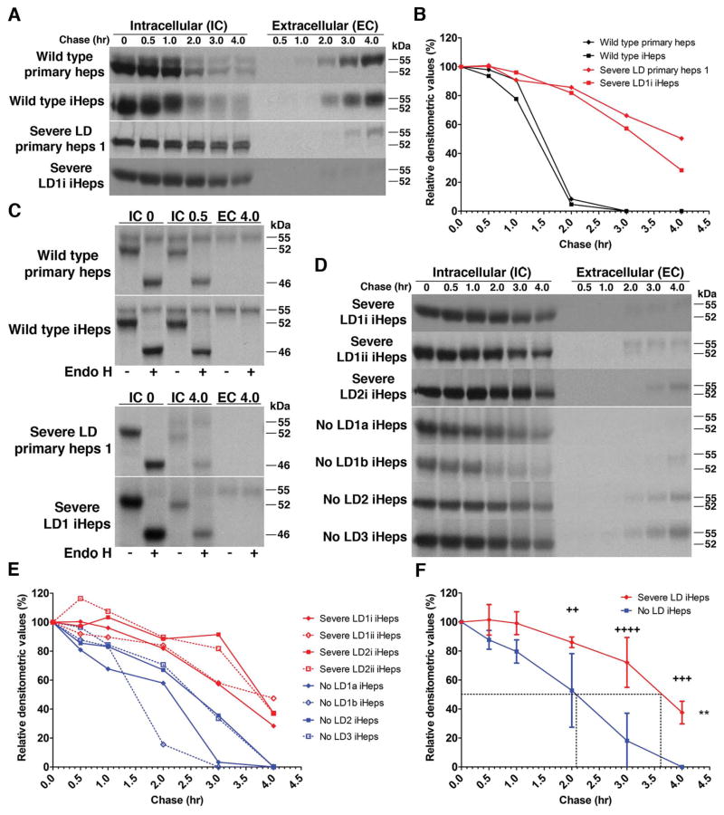
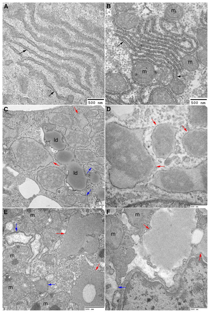
In the classical form of α1-antitrypsin deficiency (ATD), aberrant intracellular accumulation of misfolded mutant α1-antitrypsin Z (ATZ) in hepatocytes causes hepatic damage by a gain-of-function, "proteotoxic" mechanism. Whereas some ATD patients develop severe liver disease (SLD) that necessitates liver transplantation, others with the same genetic defect completely escape this clinical phenotype. We investigated whether induced pluripotent stem cells (iPSCs) from ATD individuals with or without SLD could model these personalized variations in hepatic disease phenotypes. Patient-specific iPSCs were generated from ATD patients and a control and differentiated into hepatocyte-like cells (iHeps) having many characteristics of hepatocytes. Pulse-chase and endoglycosidase H analysis demonstrate that the iHeps recapitulate the abnormal accumulation and processing of the ATZ molecule, compared to the wild-type AT molecule. Measurements of the fate of intracellular ATZ show a marked delay in the rate of ATZ degradation in iHeps from SLD patients, compared to those from no liver disease patients. Transmission electron microscopy showed dilated rough endoplasmic reticulum in iHeps from all individuals with ATD, not in controls, but globular inclusions that are partially covered with ribosomes were observed only in iHeps from individuals with SLD.

Conclusion: iHeps model the individual disease phenotypes of ATD patients with more rapid degradation of misfolded ATZ and lack of globular inclusions in cells from patients who have escaped liver disease. The results support the concept that "proteostasis" mechanisms, such as intracellular degradation pathways, play a role in observed variations in clinical phenotype and show that iPSCs can potentially be used to facilitate predictions of disease susceptibility for more precise and timely application of therapeutic strategies.


Conflict of interest statement

Potential conflict of interest: Nothing to report.

Figures

Figure 1
Figure 1
ATD iHeps recapitulate the accumulation and processing of ATZ. (A) Pulse-chase labeling showing intracellular accumulation of AT in severe LD cells but not in wild type cells. (B) Kinetics of the disappearance of AT in severe LD and wild type cells. Values are band densities of IC fractions in (A) relative to IC signal at time 0. Effect of (C) endoglycosidase H digestion on AT in pulse-chase IC and EC fractions of cells at indicated timepoints. (−) mock digestion, (+) endo H digestion. (D) Pulse-chase labeling comparing the disappearance of intracellular AT in severe and no LD iHeps. Severe LD1i and LD1ii are replicates from the same iPSc line. No LD1a and no LD1b are different iPSc clones from the same patient. (E) Kinetics of the degradation of AT in severe and no LD iHeps. Values are band densities of IC fractions in (D) relative to IC signal at time 0. (F) Composite curves of (E) shown as mean ± s.d. Dashed lines represent the half-time for disappearance of intracellular AT. **p<0.005 (two-way repeated measures ANOVA), ++p<0.01, +++p<0.001, ++++p<0.0001 (Bonferroni posttests at each time point). Although densitometric values for the no LD iHeps appear to reach zero, the absolute values are <1% in E and F.
Figure 2
Figure 2
ATZ in severe LD iHeps accumulates in rER and in compartments that are devoid of calnexin, calreticulin, or GM130. Immunofluorescent staining of severe LD iHeps for AT and calnexin (A, B), calreticulin (C, D), or GM130 (E, F). A, C, E (600X), B, D, F (2000X). (G) Single stack image of severe LD iHeps stained for AT and calnexin. (H and I) 3D surface reconstruction of multiple stacks of images of AT/calnexin-stained severe LD iHeps with calnexin signal made partially transparent to reveal AT staining inside calnexin staining. Nuclei are stained blue.
Figure 3
Figure 3
Severe LD iHeps and severe LD liver tissue sections exhibit dilated rER and globular inclusions that are partially covered with ribosomes. Electron micrograph of wild type iHeps (A), wild type liver tissue section (B), severe LD iHeps (C, D), and severe LD liver tissue section (E, F) showing normal rER (black arrows), dilated rER (blue arrows) and globular inclusions that are partially covered with ribosomes (red arrows). m: mitochondria, ld: lipid droplets.
Figure 4
Figure 4
Severe LD iHeps and no LD iHeps exhibit dilated rER but only severe LD iHeps exhibit globular inclusions. Electron micrograph of severe LD iHeps (A–C) and no LD iHeps (D–F) showing normal rER (black arrows), dilated rER (blue arrows) and globular inclusions that are partially covered with ribosomes (red arrows). ld: lipid droplets, n: nucleus, m: mitochondria.

References

    1. Chan S, Rees D. Molecular basis for the alpha1-protease inhibitor deficiency. Nature. 1975;255:240–241. - PubMed
    1. Carrell R, Jeppsson J, Laurell C, Brennan S, Owen M, Vaughan L, Boswell D. Structure and variation of human alpha 1-antitrypsin. Nature. 1982;298:329–334. - PubMed
    1. Perlmutter D, Cole F, Kilbridge P, Rossing T, Colten H. Expression of the alpha 1-proteinase inhibitor gene in human monocytes and macrophages. Proc Natl Acad Sci U S A. 1985;82:795–799. - PMC - PubMed
    1. Perlmutter DH, Silverman GA. Hepatic fibrosis and carcinogenesis in alpha1-antitrypsin deficiency: a prototype for chronic tissue damage in gain-of-function disorders. Cold Spring Harb Perspect Biol. 2011:3. - PMC - PubMed
    1. Nelson D, Teckman J, Di Bisceglie A, Brenner D. Diagnosis and management of patients with α1-antitrypsin (A1AT) deficiency. Clin Gastroenterol Hepatol. 2012;10:575–580. - PMC - PubMed

Publication types

MeSH terms

Substances