

Comparative Localization and Functional Activity of the Main Hepatobiliary Transporters in HepaRG Cells and Primary Human Hepatocytes
- PMID: 25690737
- PMCID: PMC4833040
- DOI: 10.1093/toxsci/kfv041
Comparative Localization and Functional Activity of the Main Hepatobiliary Transporters in HepaRG Cells and Primary Human Hepatocytes
Abstract
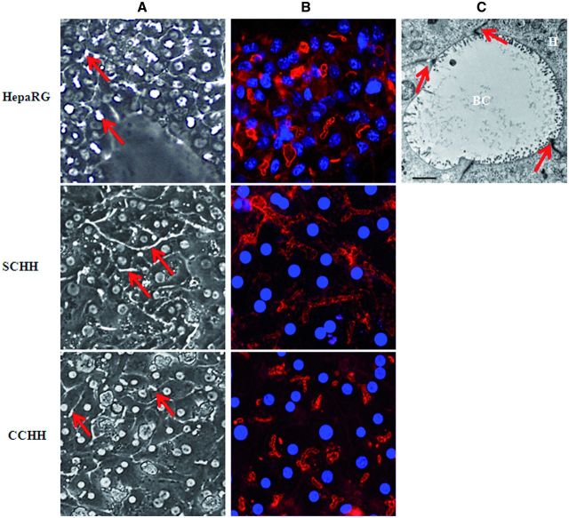
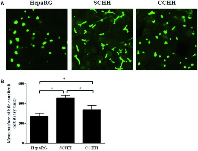
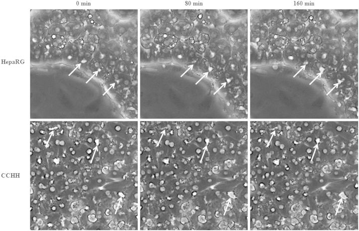
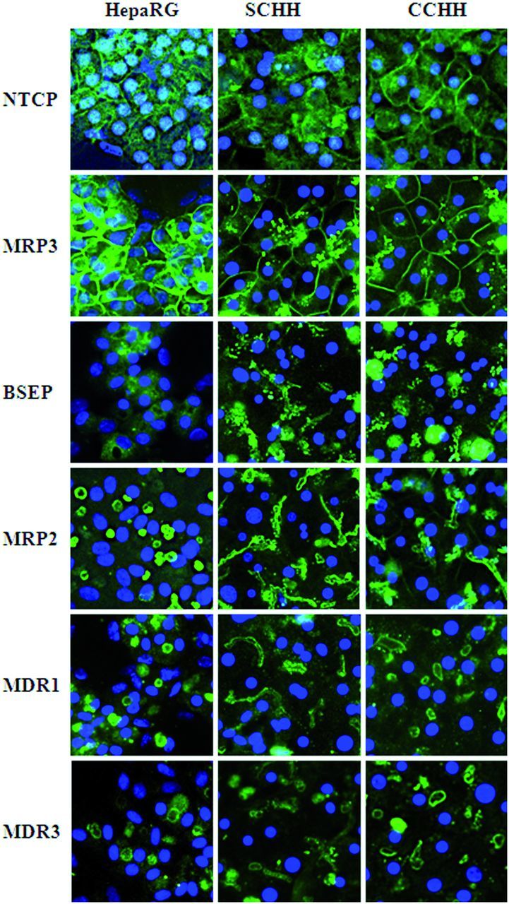
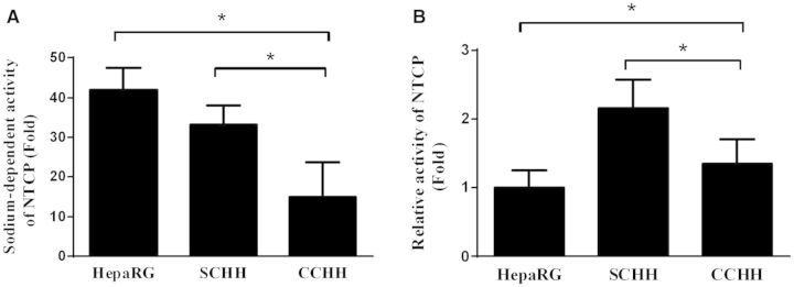
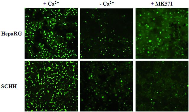
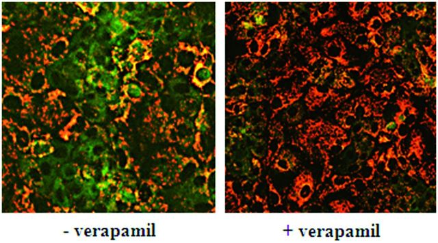
The role of hepatobiliary transporters in drug-induced liver injury remains poorly understood. Various in vivo and in vitro biological approaches are currently used for studying hepatic transporters; however, appropriate localization and functional activity of these transporters are essential for normal biliary flow and drug transport. Human hepatocytes (HHs) are considered as the most suitable in vitro cell model but erratic availability and inter-donor functional variations limit their use. In this work, we aimed to compare localization of influx and efflux transporters and their functional activity in differentiated human HepaRG hepatocytes with fresh HHs in conventional (CCHH) and sandwich (SCHH) cultures. All tested influx and efflux transporters were correctly localized to canalicular [bile salt export pump (BSEP), multidrug resistance-associated protein 2 (MRP2), multidrug resistance protein 1 (MDR1), and MDR3] or basolateral [Na(+)-taurocholate co-transporting polypeptide (NTCP) and MRP3] membrane domains and were functional in all models. Contrary to other transporters, NTCP and BSEP were less abundant and active in HepaRG cells, cellular uptake of taurocholate was 2.2- and 1.4-fold and bile excretion index 2.8- and 2.6-fold lower, than in SCHHs and CCHHs, respectively. However, when taurocholate canalicular efflux was evaluated in standard and divalent cation-free conditions in buffers or cell lysates, the difference between the three models did not exceed 9.3%. Interestingly, cell imaging showed higher bile canaliculi contraction/relaxation activity in HepaRG hepatocytes and larger bile canaliculi networks in SCHHs. Altogether, our results bring new insights in mechanisms involved in bile acids accumulation and excretion in HHs and suggest that HepaRG cells represent a suitable model for studying hepatobiliary transporters and drug-induced cholestasis.
Keywords: HepaRG hepatocytes; hepatobiliary transporters; human hepatocytes; membrane localization; transporter activity.
© The Author 2015. Published by Oxford University Press on behalf of the Society of Toxicology.All rights reserved. For Permissions, please e-mail: journals.permissions@oup.com.
Figures
References
-
- Abdel-Razzak Z., Loyer P., Fautrel A., Gautier J. C., Corcos L., Turlin B., Beaune P., Guillouzo A. (1993). Cytokines down-regulate expression of major cytochrome P-450 enzymes in adult human hepatocytes in primary culture. Mol. Pharmacol. 44, 707–715. - PubMed
-
- Antherieu S., Bachour-El Azzi P., Dumont J., Abdel-Razzak Z., Guguen-Guillouzo C., Fromenty B., Robin M. A., Guillouzo A. (2013). Oxidative stress plays a major role in chlorpromazine-induced cholestasis in human HepaRG cells. Hepatology 57, 1518–1529. - PubMed
-
- Antherieu S., Chesne C., Li R., Camus S., Lahoz A., Picazo L., Turpeinen M., Tolonen A., Uusitalo J., Guguen-Guillouzo C., et al. (2010). Stable expression, activity, and inducibility of cytochromes P450 in differentiated HepaRG cells. Drug Metab. Dispos. 38, 516–525. - PubMed
-
- Antherieu S., Chesne C., Li R., Guguen-Guillouzo C., Guillouzo A. (2012). Optimization of the HepaRG cell model for drug metabolism and toxicity studies. Toxicol. In Vitro 26, 1278–1285. - PubMed
-
- Bachour-El Azzi P., Sharanek A., Abdel-Razzak Z., Antherieu S., Al-Attrache H., Savary C. C., Lepage S., Morel I., Labbe G., Guguen-Guillouzo C., et al. (2014). Impact of inflammation on chlorpromazine-induced cytotoxicity and cholestatic features in HepaRG cells. Drug Metab. Dispos. 42, 1556–1566. - PubMed
Publication types
MeSH terms
Substances
LinkOut - more resources
Full Text Sources
Other Literature Sources
Research Materials
