. 2015 Apr 30;43(8):4013-27.
doi: 10.1093/nar/gkv056. Epub 2015 Feb 17.

The nucleoporin Mlp2 is involved in chromosomal distribution during mitosis in trypanosomatids

Affiliations

The nucleoporin Mlp2 is involved in chromosomal distribution during mitosis in trypanosomatids

Christelle Morelle et al. Nucleic Acids Res. .

Abstract

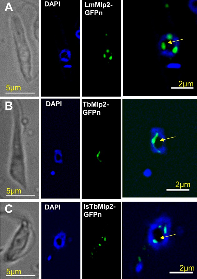
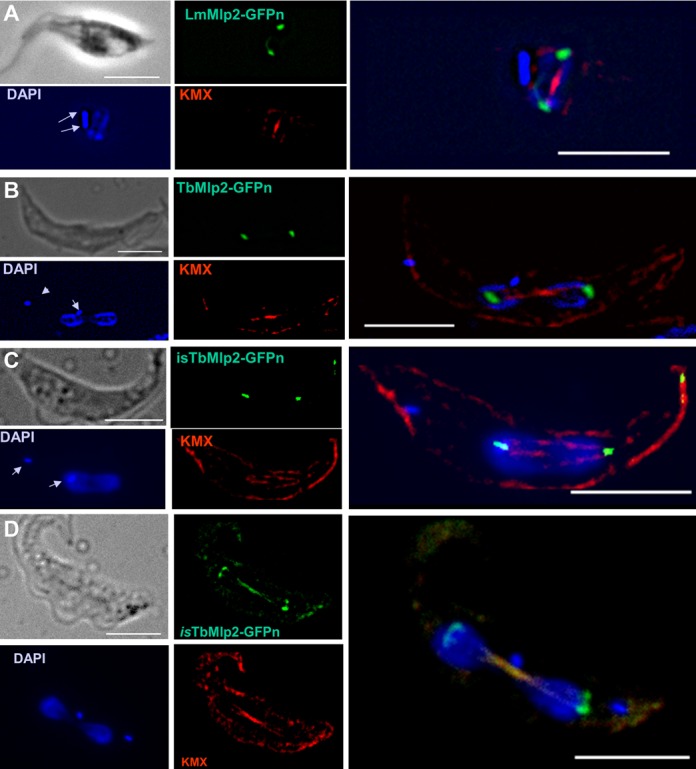
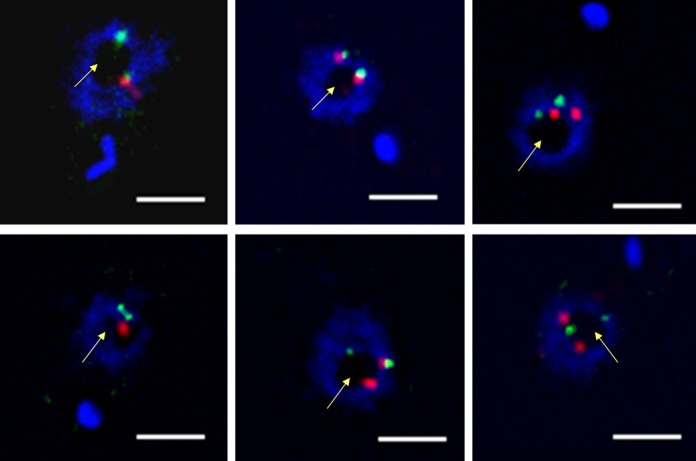
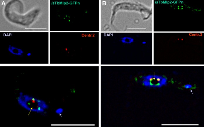
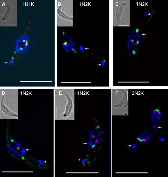
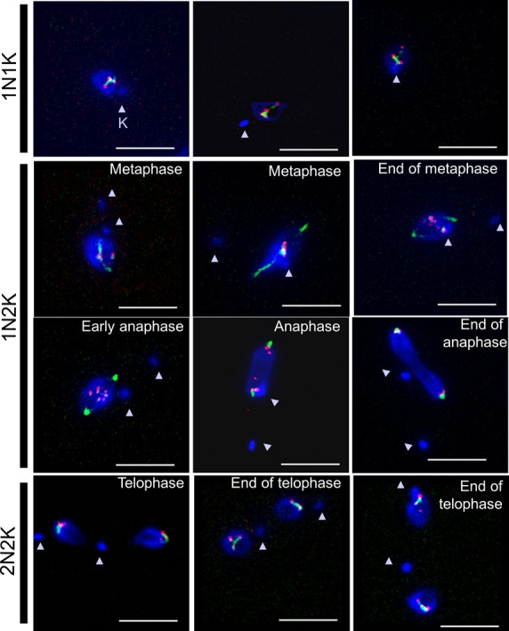
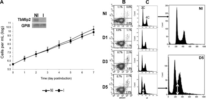
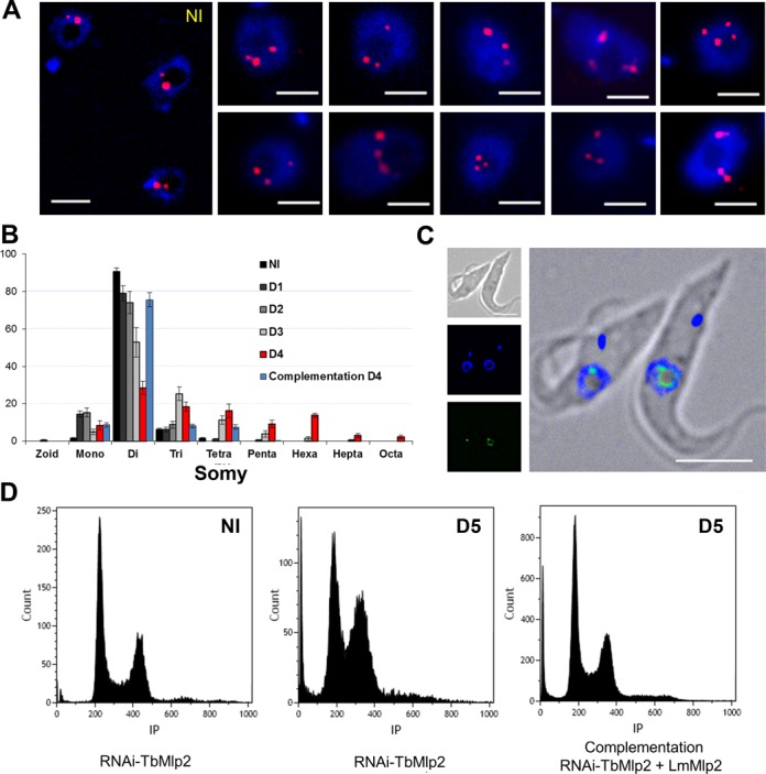
Nucleoporins are evolutionary conserved proteins mainly involved in the constitution of the nuclear pores and trafficking between the nucleus and cytoplasm, but are also increasingly viewed as main actors in chromatin dynamics and intra-nuclear mitotic events. Here, we determined the cellular localization of the nucleoporin Mlp2 in the 'divergent' eukaryotes Leishmania major and Trypanosoma brucei. In both protozoa, Mlp2 displayed an atypical localization for a nucleoporin, essentially intranuclear, and preferentially in the periphery of the nucleolus during interphase; moreover, it relocated at the mitotic spindle poles during mitosis. In T. brucei, where most centromeres have been identified, TbMlp2 was found adjacent to the centromeric sequences, as well as to a recently described unconventional kinetochore protein, in the periphery of the nucleolus, during interphase and from the end of anaphase onwards. TbMlp2 and the centromeres/kinetochores exhibited a differential migration towards the poles during mitosis. RNAi knockdown of TbMlp2 disrupted the mitotic distribution of chromosomes, leading to a surprisingly well-tolerated aneuploidy. In addition, diploidy was restored in a complementation assay where LmMlp2, the orthologue of TbMlp2 in Leishmania, was expressed in TbMlp2-RNAi-knockdown parasites. Taken together, our results demonstrate that Mlp2 is involved in the distribution of chromosomes during mitosis in trypanosomatids.


Figures

Figure 1.
Figure 1.
Subcellular localization of the nucleoporin Mlp2 in L. major and in T. brucei during interphase. (A) In L. major, LmMlp2 N-terminally fused to the GFP (LmMlp2-GFPn, green) was essentially found in the nucleus, as a limited number of more or less extended dots that localized at the periphery of the nucleolus (intranuclear area weakly labelled by DAPI, yellow arrow) (see also Supplementary Figure S1). The nuclear localization was similar for TbMlp2-GFPn in T. brucei, following either integration in the ribosomal DNA cluster (B) or in situ tagging (C). Of note, using C-terminal in situ tagging in T. brucei, a localization typical of nucleoporins could be seen in a very small proportion of cells (see Supplementary Figure S2E and F).
Figure 2.
Figure 2.
Subcellular localization of the nucleoporin Mlp2 in L. major and in T. brucei during mitosis. (A) During mitosis, LmMlp2 repositioned at the poles of the mitotic spindle, here visualized using a specific anti-tubulin antibody, KMX (red). A similar relocation was observed when the TbMlp2-GFPn recombinant gene was inserted in the ribosomal DNA cluster (B) or in situ (C, D). DAPI staining shows the duplicated (A) and then segregated kinetoplasts (B–D) (arrows) as well as dividing nuclei. Scale bar 5 μm.
Figure 3.
Figure 3.
Nuclear localization of the centromeric sequences of chromosomes 2 and 3 in T. brucei. DNA probes specific for the centromeres of chromosome 2 (green) and chromosome 3 (red) were used for FISH. These centromeric sequences were found at the periphery of the nucleolus (yellow arrow). Scale bar 2 μm.
Figure 4.
Figure 4.
Localization of TbMlp2 and centromeres of chromosomes 2 and 3 in T. brucei at interphase. In interphasic cells, the in situ tagged recombinant protein isTbMlp2-GFPn (green) and the centromeres of chromosomes 2 and 3 ((A) and (B), respectively, red) were all located at the periphery of the nucleolus (intranuclear area weakly labelled by DAPI, yellow arrow) and the centromeres found adjacent or in close proximity to TbMlp2. Bar 5 μm.
Figure 5.
Figure 5.
Dynamics of the relocation of TbMlp2 and of the centromeres during the cell cycle progress. (A) In interphasic cells (1N1K), both isTbMlp2-GFPn (green) and the centromeres of chromosome 3 (red) were found at the periphery of the nucleolus (see Figure 4). (B–D) During mitosis, which begins after the duplication of the kinetoplasts (arrow in 1N2K cells), a progressive migration of isTbMlp2-GFPn towards the mitotic spindle poles was observed. (E, F) The migration of the centromeres took place after isTb-Mlp2-GFPn had reached the pole. During their migration, centromeres were identified as two dots, but at the end of karyokinesis, the two dots were visible in each sister nucleus, materializing the two chromosome homologues. For chromosome 2, see Supplementary Figure S4. Scale bar 5 μm.
Figure 6.
Figure 6.
Dynamics of the relocation of TbMlp2 and of TbKKT4 during the cell cycle progress. The localization dynamics of one of the unconventional kinetochore proteins recently discovered in T. brucei (TbKKT4) were examined together with those of TbMlp2. First row: before the onset of mitosis (1N1K cells), both isTbMlp2-GFPn (green) and isTbKKT4-Ruby-n (red) clearly displayed an intranucleolar localization. Second row: at metaphase, TbMlp2 progressively migrated towards the spindle poles, with some of it still visible at the centre of the nucleus and metaphasic plate (right), where TbKKT4 remained. Third row: at anaphase, TbMlp2 concentrated at the spindle poles, while TbKKT4 in turn migrated to the poles. Fourth row: at telophase (2N2K cells), TbKKT4, like the centromeres (Figure 5F), was again concentrated adjacent or close to TbMlp2. Arrowheads: kinetoplasts (K). Scale bar 5 μm.
Figure 7.
Figure 7.
The RNAi-mediated knockdown of TbMlp2 induced a progressive increase of cells with DNA contents between 2C and 4C, but no cell growth reduction. (A) RNAi-mediated inhibition of TbMlp2 expression had no significant effect on cell growth. Insets: three independent RNAi experiments were performed, of which two targeted distinct sequences of TbMlp2. Northern blot controls of TbMlp2-RNAi; NI: non-induced cells. I: tetracyclin-induced cells at day 2. (B) Flow cytometry analysis: measure of phosphatidylserine exposure through the Annexin V assay after TbMlp2-RNAi. NI: non-induced cells; D1, D3, D5: tetracyclin-induced cells at day 1, day 3 and day 5 post-induction. Following depletion of TbMlp2, the exposure of phosphatidylserine to the outer membrane was not significantly altered. (C) Effect of the expression inhibition of TbMlp2 upon DNA contents in induced cells at D1, D3 and D5 versus non-induced cells. Depletion of TbMlp2 induced a progressive increase of cells with DNA contents between 2C and 4C (double white arrow).
Figure 8.
Figure 8.
Induction of aneuploidy following RNAi-mediated knockdown of TbMlp2. (A) RNA interference targeting TbMlp2 yielded variable numbers of chromosome 1 homologues (red) per nucleus. Cells on day 4 after RNAi induction were probed for FISH with an alpha–beta tubulin probe. Disomic non-induced cells (NI) are shown on the left panel. Bar: 2 μm. (B) Time evolution of chromosome 1 somy patterns observed in FISH from day 1 to day 4 after TbMlp2-RNAi, and partial restoration of diploidy at day 4 after complementation by the non-tagged LmMlp2; y-axis = cell percentages. (C) In another complementation assay using LmMlp2-GFPc, the recombinant protein was expressed in T. brucei and found at the periphery of the nucleolus. (D) Flow cytometry analysis of DNA contents after complementation with LmMlp2: in non-induced cells (NI), at Day 5 after induction, and at Day 5 after induction but with complementation with LmMlp2. Like in Figure 7C, the TbMlp2 knockdown (centre) induced an increase in cells with DNA contents between 2C and 4C as compared with non-induced cells (left). Complementation of the RNAi-knocked down parasites with LmMlp2 (right) yielded a strong shift of this phenotype towards that of the non-induced cells.

Similar articles

Cited by

References

    1. Davis L.I. The nuclear pore complex. Annu. Rev. Biochem. 1995;64:865–896. - PubMed
    1. Pante N., Aebi U. Molecular dissection of the nuclear pore complex. Crit. Rev. Biochem. Mol. Biol. 1996;31:153–199. - PubMed
    1. Gorlich D., Mattaj I.W. Nucleocytoplasmic transport. Science. 1996;271:1513–1518. - PubMed
    1. Nigg E.A. Nucleocytoplasmic transport: signals, mechanisms and regulation. Nature. 1997;386:779–787. - PubMed
    1. Rout M.P., Aitchison J.D., Suprapto A., Hjertaas K., Zhao Y., Chait B.T. The yeast nuclear pore complex: composition, architecture, and transport mechanism. J. Cell Biol. 2000;148:635–651. - PMC - PubMed

Publication types

MeSH terms

Substances

LinkOut - more resources