Review
. 2015 May;59(3):135-47.
doi: 10.1007/s10384-015-0373-5. Epub 2015 Feb 18.

Ischemic optic neuropathies and their models: disease comparisons, model strengths and weaknesses

Affiliations
Review

Ischemic optic neuropathies and their models: disease comparisons, model strengths and weaknesses

Steven L Bernstein et al. Jpn J Ophthalmol. 2015 May.

Abstract

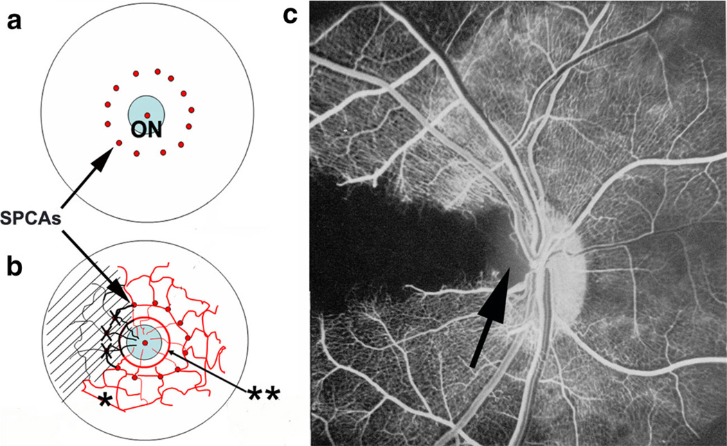
Ischemic optic neuropathies (IONs) describe a group of diseases that specifically target the optic nerve and result in sudden vision loss. These include nonarteritic and arteritic anterior ischemic optic neuropathy (NAION and AAION) and posterior ischemic optic neuropathy (NPION, APION). Until recently, little was known of the mechanisms involved in ION damage, due to a lack of information about the mechanisms associated with these diseases. This review discusses the new models that closely mimic these diseases (rodent NAION, primate NAION, rodent PION). These models have enabled closer dissection of the mechanisms involved with the pathophysiology of these disorders and enable identification of relevant mechanisms and potential pathways for effective therapeutic intervention. Descriptions of the different models are included, and comparisons between the models, their relative similarities with the clinical disease, as well as differences are discussed.


Conflict of interest statement

Conflicts of interest S. L. Bernstein, Patent (The use of Prostaglandin J2 for Optic nerve Disease); N. R. Miller, None.

Figures

Fig. 1
Fig. 1
Structure and vasculature of the human ON, and vascular schematic comparisons of the human, mouse and rat lamina and optic nerve. a Human ON structure. The ON is divided into prelaminar (intraocular), laminar and retrolaminar (postlaminar) regions. The prelaminar region (P-lam) is contiguous with the retina (Ret), and the RGC axons are unmyelinated and derive from the nerve fiber layer, with little structural delineation or restriction. Unmyelinated axon bundles are restricted by the scleral walls (Sc) in the laminar region, and separated by collagen columns. Myelination occurs in the retrolaminar region of the ON, resulting in marked enlargement of the ON diameter. Modified from [3]. b Vascular corrosion cast of the human ON regions. The laminar region (Lam) is supplied by both prelaminar (PrL-intraretinal) capillaries, as well as indirectly from the choroid, and directly from branches of the posterior ciliary arteries. The lamina vasculature is also continuous with the vascular supply of the retrolaminar (RL) ON. Capillary density of the lamina is more exuberant than the prelaminar or retrolaminar ON. Laminar blood supply is therefore more complex than any other single region. Modified from [5]. c Vascular corrosion cast of the rat ON regions. Similar to humans, the laminar capillary bed is more complex than either the pre- or retro-laminar ON regions and receives blood from retina, choroid, posterior ciliary arteries and posterior ON. Central retinal artery (A) and vein (V) are indicated. Modified from [5]. d Schematic of human ON blood supply. Vessels derived from the posterior ciliary arteries forms a plexus derived from the circle of Zinn- Haller (ZHC). The central retinal artery (CRA) derives from the ophthalmic artery and penetrates the retrolaminar ON, emerging to supply the inner retina from the center of the prelaminar (intraocular) portion of the ON. Posterior optic nerve capillaries extend from pial vessels (Pv), which themselves derive from the ophthalmic artery (OA) and vein (OV). Modified from [7]. e Schematic diagram of the mouse ON circulation. There is less complexity of the posterior ON capillaries compared with rat, but these communicate with the lamina. Retinal retrograde vasculature communicates with the lamina past the sclera (Sc), but there is reduced direct communication with the choroidal (Ch) circulation. Modified from [8]. f Schematic diagram of the rat ON circulation. Rat lamina is similar in overall structure to the primate but with reduced vascular complexity and much less supporting collagen, present mainly as plates. The posterior cilary artery (PCA) derives from the ophthalmic artery (OA) in all species. Modified from [5]
Fig. 2
Fig. 2
Optic disk appearance in a patient with unilateral NAION (a, b) and in a patient with AAION (c). a In a patient with NAION in the opposite eye, the unaffected optic disk (ON) is small, with little or no cup, and the disk margin is sharply delineated (arrowheads). b NAION-affected disk. The disk is swollen and hyperemic, with blurred disk margins (arrowheads) and obscuration of vessels (white arrow). A disk hemorrhage is present (black arrow). The veins are mildly dilated, suggesting compression at the disk. c AAION-affected disk. The ON is both pale and swollen. A cotton-wool spot is visible (arrow)
Fig. 3
Fig. 3
A schematic diagram of the primate AAION model and effect on the nonhuman primate (NHP) retina and choroid. a Schematic diagram of the normal posterior of the eye. The posterior ciliary arteries (PCAs) include the short posterior ciliary arteries (SPCAs) surrounding the optic nerve (ON) in the primate. b Primate AAION model schematic diagram. Ligation of multiple SPCAs at the entrance to the sclera results in regional choroidal ischemia, which also affects a portion of the circle of Zinn-Haller (choroidal vascular circle, in red) supplying a portion of the anterior ON, resulting in regional anterior ON ischemia. c Fluorescein retinal angiogram of a NHP immediately following regional ligation of SPCAs. The regional loss of choroidal blood flow results in compromise of the flow in the cilioretinal artery, leading to retinal capillary nonperfusion as well as nonperfusion of the underlying choroid. Regional choroidal nonperfusion has also caused loss of capillary perfusion to a portion of the ON (arrow). Photo taken from [27]
Fig. 4
Fig. 4
Rodent NAION (rNAION) model. a Control rat ON. Retinal veins are of normal caliber. The optic disk has defined margins (arrowheads) and the disk is flat against the retina. A scale bar (500 µm) indicates the diameter of the disk. b Schematic diagram of the rNAION model. The photoactive dye rose Bengal (RB) is injected, and a 500-µm-diameter laser light spot/532 nm wavelength/50 mW power is focused on the optic disc using a fundus contact lens. The laser light activates the RB, causing capillary damage. This capillary damage results in axonal ischemia. c Photo of the rat ON 1 day post-induction. The optic disk is swollen (arrowheads), with blurred margins. There is mild retinal venous dilation. d Confocal micrograph of an ON near the lamina, and overlying retina. One day post-rNAION induction, the animal was injected intravenously with a mixture of rhodamine-dye-linked 3-kDa dextran and fluoresceinlinked bovine serum (66 kDa) albumen (FITC-BSA), and euthanized 1 h later. There is accumulation of both molecules in the anterior ON with edema (asterisk) and retina overlying the nerve, suggesting localized breakdown of the blood–brain barrier (BBB) in the anterior ON and retina
Fig. 5
Fig. 5
Characteristics of the pNAION model. a Normal NHP optic disk. The ON is flat, with well defined margins. Arteries (A) and veins (V) are normal in caliber. b pNAION, 1day post-induction. The optic disk is edematous and swollen, with disk hemorrhage (arrowhead) and obscuration of vessels due to edema (arrow). c pNAION, 30days post-induction. There is temporal pallor of the affected nerve (indicated by arrows). d Fluorescein angiogram (late phase) of the pNAION-induced (1 day) ON. There is disk leakage from the ischemic regions (arrowheads). e SD-OCT of an pNAION-affected eye, 1day post-induction. The ON is swollen, and subretinal fluid is apparent in the macula (indicated by arrows). Mac: macula. f SDOCT ring scan of retina shown in e. The RNFL is regionally swollen, and there is subretinal fluid (SRF) apparent in the region of the macula. g Histological (H&E stained) section of a pNAION lamina and ON, 70 days post-induction. There is regional axonal loss (indicated by arrows) and increased cellularity in the loss region, consistent with inflammatory cell infiltrate
Fig. 6
Fig. 6
Rodent PION model. a photograph of the laser set-up. Light from a continuous wave laser is segmented by a ‘chopper’, further processed and filtered, then directed inferiorly using a mirror. b The ON is exposed posteriorly to the globe. c The photoreactive dye (erythrosin B), is activated by a laser spot placed on the surface of the ON. d Post-illumination, the treated nerve exhibits vascular damage and surface hemorrhage (arrow). e. Disk photo of the untreated ON. f Disk photo of the PIONinduced ON 4 days posttreatment. There is no observable disk edema. g Disk photo of a PION-induced eye 28 days post-induction. The optic disk is slightly shrunken in appearance and exhibits slight pallor. Modified from [43]
Fig. 7
Fig. 7
ON ischemic stress results in regional RGC stress in the mouse after rNAION. A transgenic mouse line was generated with a c-Fos-driven promotor-reporter gene construct (cfos-lacZ) [74]. Three days following induction, the whole retina was immunostained for lacZ expression. a Region 1 reveals low levels of lacZ protein (arrow) in the RGC layer. b Region 2 reveals that nearly all RGCs express high levels of cfos-driven lacZ (arrow). Scale bar in b 50 microns

References

    1. Arnold AC. Ischemic optic neuropathies. Ophthalmol Clin North Am. 2001;14:83–98. - PubMed
    1. Hayreh SS. Posterior ischaemic optic neuropathy: clinical features, pathogenesis, and management. Eye (Lond) 1969;18:1188–1206. - PubMed
    1. Anderson DR. Ultrastructure of human and monkey lamina cribrosa and optic nerve head. Arch Ophthalmol. 1969;82:800–814. - PubMed
    1. Cioffi GA, Van Buskirk EM. Microvasculature of the anterior optic nerve. Surv Ophthalmol. 1994;38(Suppl):S107. - PubMed
    1. Morrison JC, Johnson EC, Cepurna WO, Funk RH. Microvasculature of the rat optic nerve head. Invest Ophthalmol Vis Sci. 1999;1999(40):1702–1709. - PubMed

Publication types

LinkOut - more resources