Review
. 2015 Mar;20(2):250-63.
doi: 10.1007/s00776-014-0687-z. Epub 2015 Feb 19.

Diagnosis and treatment of Ewing sarcoma of the bone: a review article

Affiliations
Review

Diagnosis and treatment of Ewing sarcoma of the bone: a review article

Toshifumi Ozaki. J Orthop Sci. 2015 Mar.

Abstract

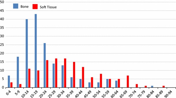
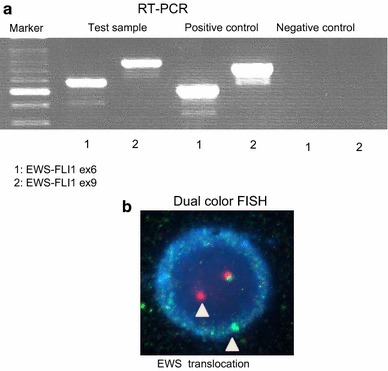
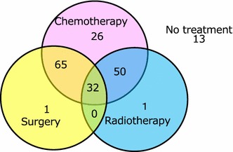
Ewing sarcoma (ES) is rare in Japanese people, and only 30-40 patients develop the disease annually. To diagnose ES, molecular techniques that aim to detect characteristic fusion genes are commonly used in combination with conventional histological and immunohistochemical examinations. The treatment strategy for ES is characterized by multi-disciplinary collaboration between pediatric oncologists, medical oncologists, radiation oncologists, and orthopedic surgeons. In recent years, numerous large-scale national or international multi-institutional studies of ES have been performed. Pre- and postoperative intensive systemic chemotherapy with multiple anticancer drugs is the standard treatment method for ES. Depending on the obtained surgical margin, postoperative radiation might also be performed. If preoperative radiological examinations indicate that surgical excision would be difficult, preoperative radiation can be administered. As the treatment outcomes of ES have improved, late complications and secondary malignancies have become a problem. After treatment, patients with ES require very long-term follow-up in order to detect secondary malignancies and growth-related musculoskeletal complications.


Figures

Fig. 1
Fig. 1
The age distribution of patients with ES of the bone (blue bar) and extraskeletal ES (red bar) [27, 28]
Fig. 2
Fig. 2
A 38-year-old woman with ES of the right tibia. a Plain X-ray showing permeative bone destruction. MRI detected a tumor that displayed b, low signal intensity and c, heterogeneous high signal intensity on T1- and T2-weighted imaging, respectively. The tumor was located both around and within the tibia. This case was presented by Dr Akira Kawai, Department of Musculoskeletal Oncology and Rehabilitation, National Cancer Center Hospital, Tokyo, Japan
Fig. 3
Fig. 3
A 16-year-old patient with ES of the ilium. a A radiolucent lesion was visible in the right ilium on a plain X-ray (arrowhead). b CT detected a large mass that exhibited a periosteal reaction in the right ilium (arrowhead) and c, T2-weighted MRI depicted a mass that displayed heterogeneous high signal intensity in and around the right ilium and acetabulum
Fig. 4
Fig. 4
A 9-year-old girl with ES developed a metastasis in her left ischium. a T2-weighted MRI (T2-STIR) demonstrated an area of high signal intensity in the ischium, and 18F-FDG-PET detected high 18F-FDG uptake in the same region
Fig. 5
Fig. 5
Histology of ES. a ES involves the sheet-like proliferation of small round cells with little cytoplasm (hematoxylin-eosin stain). b Periodic acid-Schiff staining detected small positive granules, which were considered to be glycogen molecules
Fig. 6
Fig. 6
Membranous expression of CD99, a product of the MIC2 gene, is observed in ES cells
Fig. 7
Fig. 7
a RT-PCR. The left lane shows a marker, and the next three pairs of lanes (in a left-to-right direction) show the test samples, positive controls, and negative controls (1: EWS-FLI1 ex6, 2: EWS-FLI1 ex9), respectively. This figure shows that the test sample was positive for both EWS-FLI1 ex6 and EWS-FLI1 ex9, which means that the tumor possessed the EWS-FLI1 ex6 fusion gene. b FISH. An abnormal cell that has been hybridized with the Vysis LSI EWSR1 (22q12) dual color, break apart rearrangement probe. In a normal cell that lacks a t(22q12) translocation in the EWSR1 gene region, two fusion signals will be observed, reflecting the presence of two intact copies of EWSR1. The cell in this image shows one fusion, one orange, and one green signal, which is indicative of the rearrangement of one copy of the EWSR1 gene region
Fig. 8
Fig. 8
Treatments employed in 188 cases of ES (2006–2011). The data shown are derived from the Bone Tumor Registry in Japan, 2010, run by the JOA Musculoskeletal Tumor Committee
Fig. 9
Fig. 9
An 18-year-old girl had a 1-month history of back pain without any obvious cause and experienced muscle weakness in both legs The patient suffered numbness and urinary disturbances. a Plain X-ray showing the loss of spinous processes at the T5 level (arrow). b CT detected a spotted osteolytic lesion in the lamina of the T5 vertebra. c T2-weighted MRI showed a mass that exhibited iso-signal intensity and was compressing the spinal cord from the posterior direction (arrow). d Emergent decompression of the T4-6 vertebrae was performed to rescue the spinal cord from compression. Preoperative chemotherapy composed of VDC + IE was performed, 45 Gy radiation was administered as a local treatment, and VDC-IE was administered as a post-local therapy. The patient became completely free from spinal palsy and was able to walk without any support. She has been disease-free for 6 years after the local treatment
Fig. 10
Fig. 10
A 10-year-old boy with ES of the right humerus. He had a pain in his right upper arm. A plain X-ray showed a limited periosteal reaction. a T1-weighted MRI depicted an area of heterogeneous low signal intensity in the humerus. b A bone scan showed abnormally high isotope uptake in the right humerus. After the diagnosis of ES had been confirmed by open biopsy, preoperative chemotherapy composed of DOX, CPM, methotrexate, VCR, IFO, and ACT-D was administered. c After the preoperative chemotherapy, the patient underwent wide excision of the tumor in his right humerus. A sling procedure involving a vascularized fibula graft was performed for reconstruction, and postoperative chemotherapy was initiated. At 13 years after surgery, he visited our hospital due to dyspnea and coughing. A chest X-ray showed a white region in his right lung, and d CT depicted a large amount of pleural effusion and atelectasis due to the pulmonary metastasis of ES
Fig. 11
Fig. 11
A 1-year-old boy suddenly suffered a pain in his right lower leg without any obvious cause. a A plain X-ray showed a radiolucent lesion and widening of the diaphysis of his right tibia. A periosteal reaction was also observed. b T2-weighted imaging demonstrated an area of high signal intensity in the tibia. An open biopsy resulted in a diagnosis of ES of the tibia. After preoperative chemotherapy composed of 6 courses of VIDE according to the protocol outlined in the Euro-EWING 99 study, radiotherapy involving 55.8 Gy was performed as a local therapy. After the radiotherapy, one course of the VAI regimen and 7 courses of the VAC regimen were administered. When the patient was 9.5 years old, CT detected lymph node swelling in his neck. A biopsy of the lymph node demonstrated metastasis from papillary thyroid cancer. The patient underwent excision of his right thyroid lobe and the swollen lymph node. c He displayed a 5-cm leg length discrepancy at 10 years after the local therapy

References

    1. Jaffe R, Santamaria M, Yunis EJ, Tannery NH, Agostini RM, Jr, Medina J, Goodman M. The neuroectodermal tumor of bone. Am J Surg Pathol. 1984;8(12):885–898. doi: 10.1097/00000478-198412000-00001. - DOI - PubMed
    1. Carlei F, Polak JM, Ceccamea A, Marangos PJ, Dahl D, Cocchia D, Michetti F, Lezoche E, Speranza V. Neuronal and glial markers in tumours of neuroblastic origin. Virchows Arch A Pathol Anat Histopathol. 1984;404(3):313–324. doi: 10.1007/BF00694896. - DOI - PubMed
    1. Hashimoto H, Enjoji M, Nakajima T, Kiryu H, Daimaru Y. Malignant neuroepithelioma (peripheral neuroblastoma). A clinicopathologic study of 15 cases. Am J Surg Pathol. 1983;7(4):309–318. doi: 10.1097/00000478-198306000-00002. - DOI - PubMed
    1. de Alava E, Lessnick SL, Sorensen PH. Ewing sarcoma. In: Fletcher CDM, Bridge JA, Hogendoorn PCW, Mertens F, editors. WHO classification of tumours of soft tissue and bone. Lyon: WHO Press; 2013. pp. 305–309.
    1. Delattre O, Zucman J, Plougastel B, Desmaze C, Melot T, Peter M, Kovar H, Joubert I, de Jong P, Rouleau G, Aurias A, Thomas G. Gene fusion with an ETS DNA-binding domain caused by chromosome translocation in human tumours. Nature. 1992;359(6391):162–165. doi: 10.1038/359162a0. - DOI - PubMed

Publication types