. 2015 Feb 17;6(1):e02068-14.
doi: 10.1128/mBio.02068-14.

E6^E7, a novel splice isoform protein of human papillomavirus 16, stabilizes viral E6 and E7 oncoproteins via HSP90 and GRP78

Affiliations

E6^E7, a novel splice isoform protein of human papillomavirus 16, stabilizes viral E6 and E7 oncoproteins via HSP90 and GRP78

Masahiko Ajiro et al. mBio. .

Abstract

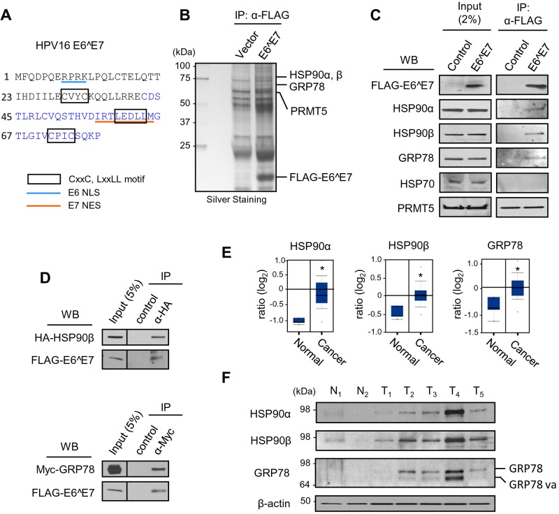
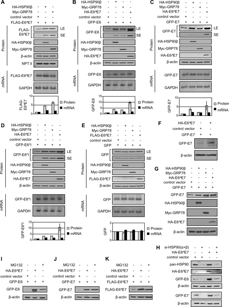
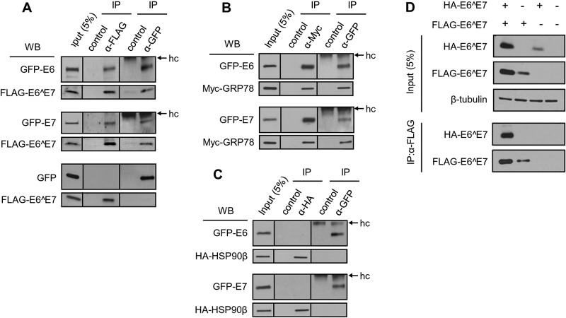
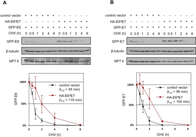
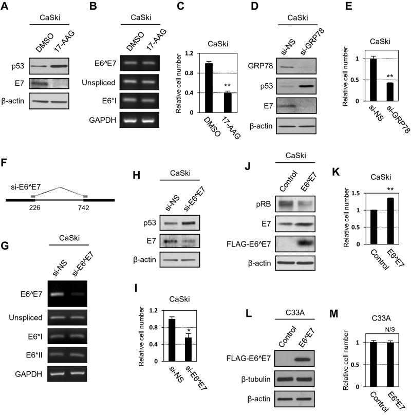
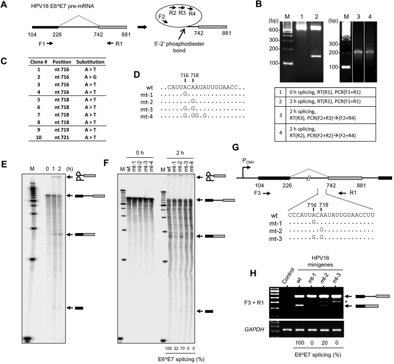
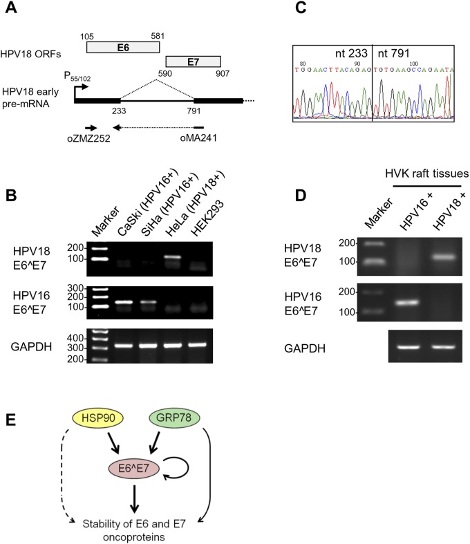
Transcripts of human papillomavirus 16 (HPV16) E6 and E7 oncogenes undergo alternative RNA splicing to produce multiple splice isoforms. However, the importance of these splice isoforms is poorly understood. Here we report a critical role of E6^E7, a novel isoform containing the 41 N-terminal amino acid (aa) residues of E6 and the 38 C-terminal aa residues of E7, in the regulation of E6 and E7 stability. Through mass spectrometric analysis, we identified that HSP90 and GRP78, which are frequently upregulated in cervical cancer tissues, are two E6^E7-interacting proteins responsible for the stability and function of E6^E7, E6, and E7. Although GRP78 and HSP90 do not bind each other, GRP78, but not HSP90, interacts with E6 and E7. E6^E7 protein, in addition to self-binding, interacts with E6 and E7 in the presence of GRP78 and HSP90, leading to the stabilization of E6 and E7 by prolonging the half-life of each protein. Knocking down E6^E7 expression in HPV16-positive CaSki cells by a splice junction-specific small interfering RNA (siRNA) destabilizes E6 and E7 and prevents cell growth. The same is true for the cells with a GRP78 knockdown or in the presence of an HSP90 inhibitor. Moreover, mapping and alignment analyses for splicing elements in 36 alpha-HPVs (α-HPVs) suggest the possible expression of E6^E7 mostly by other oncogenic or possibly oncogenic α-HPVs (HPV18, -30, -31, -39, -42, -45, -56, -59, -70, and -73). HPV18 E6^E7 is detectable in HPV18-positive HeLa cells and HPV18-infected raft tissues. All together, our data indicate that viral E6^E7 and cellular GRP78 or HSP90 might be novel targets for cervical cancer therapy.

Importance: HPV16 is the most prevalent HPV genotype, being responsible for 60% of invasive cervical cancer cases worldwide. What makes HPV16 so potent in the development of cervical cancer remains a mystery. We discovered in this study that, besides producing two well-known oncoproteins, E6 and E7, seen in other high-risk HPVs, HPV16 produces E6^E7, a novel splice isoform of E6 and E7. E6^E7, in addition to self-interacting, binds cellular chaperone proteins, HSP90 and GRP78, and viral E6 and E7 to increase the steady-state levels and half-lives of viral oncoproteins, leading to cell proliferation. The splicing cis elements in the regulation of HPV16 E6^E7 production are highly conserved in 11 oncogenic or possibly oncogenic HPVs, and we confirmed the production of HPV18 E6^E7 in HPV18-infected cells. This study provides new insight into the mechanism of splicing, the interplay between different products of the polycistronic viral message, and the role of the host chaperones as they function.


Figures

FIG 1
FIG 1
Coimmunoprecipitation (co-IP) and LC-MS/MS analysis identified HSP90α, HSP90β, and GRP78 as E6^E7-interacting proteins. (A) Diagram of HPV16 E6^E7 amino acid residues. E6- and E7-derived amino acid residues are indicated in black and blue letters, respectively. The E6-derived nuclear localization signal (NLS) and E7-derived nuclear export signal (NES) are underlined. CXXC and LXXLL motifs are boxed. (B) Silver staining image of anti-FLAG-immunoprecipitated (α-FLAG) proteins from HEK293 cells transfected with a FLAG-E6^E7 expression vector or an empty vector for 48 h. Proteins identified by LC-MS/MS are indicated on the right. (C and D) Western blot (WB) verification of the specific binding of endogenous HSP90α, HSP90β, and GRP78, but not HSP70 and PRMT5, to FLAG-E6^E7 by anti-FLAG IP (C) or the binding of ectopic HA-HSP90β or Myc-GRP78 to FLAG-E6^E7 by anti-HA IP (D, top panel) or anti-Myc IP (D, lower panel). Cell lysate from an empty vector transfection served as an IP control in panel C, and Sepharose beads without antibody were used as an IP control in panel D. HEK293 cell lysates prepared at 48 h of transfection were used in all IP and Western blot assays. Western blotting was conducted with an antibody specific for an endogenous protein or a tag-specific antibody for an ectopic protein tag. (E) Relative mRNA levels of HSP90α, HSP90β, and GRP78 in normal cervix and cervical cancer tissues. *, P < 1 × 10⁻⁴ (Student’s t test between two groups). Normal cervix specimens, n = 5; cervical cancer tissue specimens, n = 40. (F) Western blot analysis of HSP90α, HSP90β, GRP78, and GRP78va (a GRP78 splice isoform) (31) for normal cervix specimens (lanes N1 and N2) and cervical cancer tissues (lanes T1, T2, T3, T4, and T5). β-actin was used as an internal loading control.
FIG 2
FIG 2
HPV16 E6^E7, HSP90, and GRP78 promote the protein stability of HPV16 E6 and E7. (A to E) ΗSP90β and GRP78 increase the protein but not mRNA levels of HPV16 E6^E7, E6, and E7 in HEK293 cells. Following cotransfection with the indicated vectors in each panel for 48 h, HEK293 cells were analyzed by Western blotting for protein expression levels of FLAG-E6^E7 (A), GFP-E6 (B), GFP-E7 (C), GFP-E6*I (D), or GFP (E) and by Northern blotting for the expression levels of corresponding mRNAs. An empty vector without any tagged protein expression served as a control in each transfection, and neomycin phosphotransferase II (NPT II, a neomycin resistance gene product) served as a control for plasmid transfection and expression efficiency. LE, longer exposure; SE, shorter exposure. After normalization with β-actin in a Western blot or with GAPDH in a Northern blot, the relative (fold) changes in the levels of the corresponding protein or mRNA in HEK293 cells cotransfected with HA-HSP90β, Myc-GRP78, or HA-E6^E7 over the levels in cells transfected with an empty control vector are shown at the bottom of each panel in bar graphs. Error bars indicate standard deviations from two different blots. (F) E6^7 increases the E7 protein level in primary human foreskin keratinocytes. The keratinocytes were cotransfected with 4D-Nucleofector and GFP-E6 plus an HA-E6^E7 expression vector or an empty control vector for 48 h and analyzed by Western blotting. (G) HA-HSP90β, Myc-GRP78, and HA-E6^E7 increase GFP-E7 protein level in HeLa cells. HeLa cells were cotransfected with the indicated vectors for 48 h and analyzed by Western blotting. (H) The effect of HPV16 E6^E7 on E6 and E7 stability relies on HSP90. HEK293 cells were transfected twice, with a 48-h interval, with siRNAs specific for HSP90α and -β isoforms or a nontargeting control siRNA (−) for 4 days. During the second siRNA transfection, the cells were cotransfected with an HA-E6^E7 expression vector or a control vector (p3×FLAG-CMV14) in combination with a GFP-E6 or -E7 expression vector for 24 h and analyzed by Western blotting for HSP90α and -β knockdown efficiency with a pan-HSP90 antibody, E6^E7 with an anti-HA antibody, or HPV16 E6 or E7 with an anti-GFP antibody. β-actin served as a loading control. (I to K) HA-E6^E7 increased protein levels, and the levels of MG132-stabilized GFP-E6, GFP-E7, and FLAG-E6^E7 are comparable in HEK293 cells at 48 h of transfection. The cell cotransfections were conducted with a GFP-E6 (I), GFP-E7 (J), or FLAG-E6^E7 (K) expression vector along with an HA-E6^E7 expression vector or control vector. For MG132 treatment, the cells transfected with a GFP-E6, GFP-E7, or FLAG-E6^E7 expression vector were treated with MG132 (10 µM) or an equivalent amount of dimethyl sulfoxide (DMSO) 6 h prior to being harvested for Western blotting with an anti-GFP or anti-FLAG antibody. β-actin served as a sample loading control.
FIG 3
FIG 3
HPV16 E6 and E7 oncoproteins are proteins that interact with E6^E7 and GRP78. (A to C) HPV16 E6 and E7 interact with E6^E7 (A) and GRP78 (B), but not with HSP90β(C). HEK293 cells were transfected with FLAG-E6^E7 (A), Myc-GRP78 (B), or HA-HSP90β (C) along with GFP-E6 or -E7 (A to C) for 48 h. The cell lysates were immunoprecipitated with the corresponding antibody, as indicated. Rabbit IgG was used as a negative control for each anti-GFP IP, and Sepharose beads without antibody served as controls for anti-FLAG, anti-HA, and anti-Myc IP. The interacting proteins in coimmunoprecipitations or in the input were examined by Western blotting (WB) with anti-GFP for GFP-E6 or -E7 (A to C), anti-FLAG for FLAG-E6^E7 (A), anti-Myc for Myc-GRP78 (B), or anti-HA antibody for HA-HSP90β(C). hc, IgG heavy chain. (D) E6^E7 is a self-interacting protein. HEK293 cells were cotransfected with a FLAG-E6^E7 and an HA-E6^E7 expression vector or an empty (−) control vector for 48 h. The cell lysates were blotted for the expression level of each protein (input panel) and immunoprecipitated with anti-FLAG antibody. The proteins pulled down by IP were blotted with an anti-FLAG antibody for FLAG-E6^E7 or anti-HA antibody for HA-E6^E7. β-Tubulin served as a sample loading control.
FIG 4
FIG 4
HPV16 E6^E7 stabilizes E6 and E7 and prolongs the protein half-life. HEK293 cells were cotransfected with a GFP-E6 (A) or GFP-E7 (B) expression vector together with an HA-E6^E7 or an empty control vector. After 16 h of cotransfection, the cells were treated with 0.1 mg/ml of cycloheximide (CHX) for the indicated times (0, 0.5, 1, 2, 4, and 8 h) before the sample collection for Western blotting with an anti-GFP antibody, anti-β-tubulin for sample loading, or anti-NPT II antibody for plasmid transfection efficiency. The half-life (t1/2) of GFP-E6 or GFP-E7 was determined from a line plot analysis according to the following formulas, with its expression level at time zero being set to 100% and the two lines (x, y) crossing the 50% decay point (y + 0.5): for GFP-E6 with the control vector, y = −0.9583x + 1.2217; for GFP-E6 with HA-E6^E7, y = −0.3683 + 1.2291; for GFP-E7 with the control vector, y = −0.6238x + 1.0828; and for GFP-E7 with HA-E6^E7, y = −0.2802x + 1.2179, where x is the CHX treatment time (h) and y is the relative protein expression levels of GFP-E6 (A) or GFP-E7 (B). Black squares, protein expression level of GFP-E6 (A) or GFP-E7 (B) in HEK293 cells cotransfected with a control vector; red squares, protein expression level of GFP-E6 (A) or GFP-E7 (B) in HEK293 cells cotransfected with an HA-E6^E7 expression vector.
FIG 5
FIG 5
HSP90, GRP78, and E6^E7 are required to maintain a steady-state level of E6 and E7 in HPV16-positive CaSki cells. (A to C) A functional HSP90 is required for the stability of HPV16 E6 and E7 and proliferation of cervical cancer cells. HPV16-positive CaSki cells treated with 5 µM 17-AAG or DMSO for 48 h were examined by Western blotting (A), RT-PCR (B), and a cell proliferation assay (C). p53 was used to indicate E6. GAPDH served as a loading control in RT-PCR. (D and E) Knockdown of GRP78 expression in CaSki cells destabilizes E6 and E7 and inhibits cell growth. CaSki cells treated twice with 40 nM nontargeting siRNA (si-NS) or GRP78 siRNA (si-GRP78) for 96 h were examined by Western blotting (D) and a cell proliferation assay (E). (F to I) Knockdown of E6^E7 expression in CaSki cells destabilizes viral E6 and E7 and prevents cell growth. An E6^E7-specific siRNA (si-E6^E7) for the nt 226-to-nt 742 splice junction (F) was designed to silence E6^E7 expression without affecting other E6 splice isoform RNAs, as shown by RT-PCR (G). CaSki cells transfected twice with 40 nM si-NS or si-E6^E7 at 48-h intervals for 96 h were examined for E6 (p53) and E7 expression (H) and cell proliferation (I). See the details in panels A to C. (J and K) Overexpression of E6^E7 in CaSki cells increases E7 stability (J) and promotes cell proliferation (K). CaSki cells were transfected twice (for Western blotting at day 5) or three times (for cell proliferation at day 7) with a FLAG-E6^E7 or an empty vector at 24-h intervals. (L and M) Overexpression of E6^E7 has no effect on C33A, an HPV-negative cervical cancer cell line containing mutations in both p53 and pRB. C33A cells transfected with a FLAG-E6^E7 or an empty vector as described for panels J and K served as a cell line control to CaSki cells, and results were analyzed by Western blotting (L) and a cell proliferation assay (M) at days 5 and 7, respectively. *, P < 0.05; **, P < 0.01. Nonsignificance (N/S), P ≥ 0.05 by Student’s t test (C and E, I and K, and M). (A, D, H, J, and L) β-Actin served as a sample loading control.
FIG 6
FIG 6
Mapping of RNA cis elements for splicing of HPV16 E6^E7 pre-mRNA. (A) Diagram of a lariat RT-PCR strategy to map the BPS responsible for splicing of E6^E7 pre-mRNA, with the indicated primers (F1 to F2 and R1 to R4). (B) Validation of in vitro splicing (2 h) of E6^E7 pre-mRNA by RT-PCR. The left panel indicates the fully spliced products at a size of 136 nt. The right panel indicates the products of lariat RT-PCR at a size of ~200 nt. Combinations of primers used in RT-PCR are indicated in the box below. Lane M, molecular size markers. (C) Summary of the mapped branch sites by lariat RT-PCR. (D) Introduction of the A-to-G mutation (mt-1, mt-2, mt-3, and mt-4) into the mapped branch sites for in vitro RNA splicing. Nucleotides identical to the wild-type (wt) nucleotide are indicated by dots. (E) Reconstitution of E6^E7 RNA splicing in vitro. Following the splicing reaction in HeLa cell nuclear extract for the indicated times (h), the spliced products were analyzed in a 6% polyacrylamide gel with 7.5 M urea. Identities of each band are indicated on the right. (F) E6^E7 pre-mRNAs with wt or mutant branch sites were used for a 2-h reaction of in vitro splicing. Relative E6^E7 splicing efficiencies (percentages) were calculated as described previously (12) and are indicated in the bottom. The identities of each band are indicated at the right. (G) Diagram of the minigene vectors to express E6^E7 pre-mRNAs with a wt or mutant branch site (mt-1, mt-2, or mt-3). (H) RT-PCR was performed on total RNA of HEK293 cells transfected with E6^E7 minigenes or a control vector for 24 h to detect unspliced and spliced E6^E7 mRNAs (for the upper panel, primers F3 and R1 were used). GAPDH served as a loading control (lower panel). The identity of each band is indicated at the right. *, a nonspecific amplicon. Relative E6^E7 splicing efficiencies (percentages) are indicated at the bottom.
FIG 7
FIG 7
Expression of HPV18 E6^E7 in an HPV18-infected cell line and raft tissues. (A) Diagram of alternative RNA splicing from the nt 233 5′ ss to the nt 791 3′ ss to produce E6^E7 mRNA from HPV18 early transcripts. The ORFs of the HPV18 E6 and E7 oncogenes are indicated at the top. Primers specific for HPV18 E6^E7 mRNA detection by RT-PCR are indicated below the pre-mRNA. (B) Detection of HPV18 E6^E7 from HPV18-positive HeLa cells and HPV16 E6^E7 from HPV16-positive CaSki or SiHa cells by RT-PCR. HPV-negative HEK293 cells and GAPDH mRNA served as controls. (C) Determination of the HPV18 E6^E7 splice junction by sequencing of RT-PCR products gel purified from the experiment whose results are shown in panel B. (D) Detection of HPV18 E6^E7 and HPV16 E6^E7 in the HVK raft tissues with the corresponding virus infection. (E) E6^E7 plays a central role in HSP90 and GRP78 regulation of HPV16 E6 and E7 protein stability through protein-protein interactions. HSP90 and GRP78 are also involved in the stability of HPV16 E6 and E7, either indirectly (dashed arrow) or directly (solid arrow).

References

    1. Bernard HU, Burk RD, Chen Z, van Doorslaer K, zur Hausen H, de Villiers EM. 2010. Classification of papillomaviruses (PVs) based on 189 PV types and proposal of taxonomic amendments. Virology 401:70–79. doi:10.1016/j.virol.2010.02.002. - DOI - PMC - PubMed
    1. Howley PM, Lowy DR. 2007. Papillomaviruses, p 2299–2354. In Knipe DM, Howley PM, Griffin DE, Lamb RA, Martin MA, Roizman B, Straus SE (ed), Fields virology, 5th ed. Lippincott Williams & Wilkins, Philadelphia, PA.
    1. Walboomers JM, Jacobs MV, Manos MM, Bosch FX, Kummer JA, Shah KV, Snijders PJ, Peto J, Meijer CJ, Muñoz N. 1999. Human papillomavirus is a necessary cause of invasive cervical cancer worldwide. J Pathol 189:12–19. doi:10.1002/(SICI)1096-9896(199909)189:1<12::AID-PATH431>3.0.CO;2-F. - DOI - PubMed
    1. Muñoz N, Bosch FX, de Sanjosé S, Herrero R, Castellsagué X, Shah KV, Snijders PJ, Meijer CJ, International Agency for Research on Cancer Multicenter Cervical Cancer Study Group . 2003. Epidemiologic classification of human papillomavirus types associated with cervical cancer. N Engl J Med 348:518–527. doi:10.1056/NEJMoa021641. - DOI - PubMed
    1. Munoz N, Castellsague X, de Gonzalez AB, Gissmann L. 2006. Chapter 1: HPV in the etiology of human cancer. Vaccine 24(Suppl 3):S3/1–S3/10. doi:10.1016/j.vaccine.2006.05.115. - DOI - PubMed

MeSH terms

Substances

LinkOut - more resources