. 2015 Feb 17;6(1):e02357-14.
doi: 10.1128/mBio.02357-14.

Novel components of the Toxoplasma inner membrane complex revealed by BioID

Affiliations

Novel components of the Toxoplasma inner membrane complex revealed by BioID

Allan L Chen et al. mBio. .

Abstract

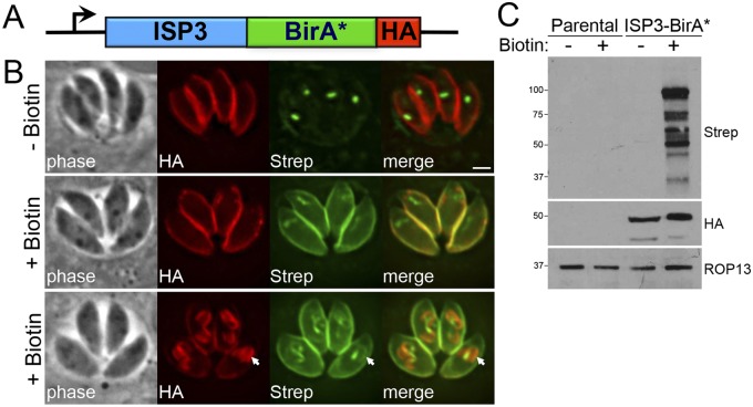
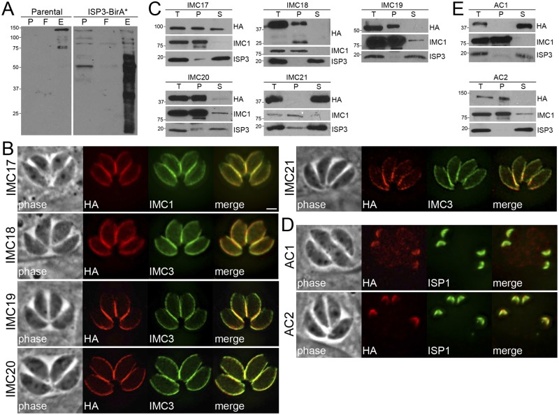
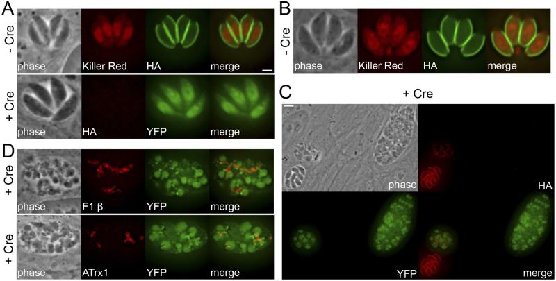
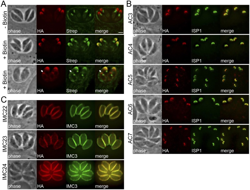
The inner membrane complex (IMC) of Toxoplasma gondii is a peripheral membrane system that is composed of flattened alveolar sacs that underlie the plasma membrane, coupled to a supporting cytoskeletal network. The IMC plays important roles in parasite replication, motility, and host cell invasion. Despite these central roles in the biology of the parasite, the proteins that constitute the IMC are largely unknown. In this study, we have adapted a technique named proximity-dependent biotin identification (BioID) for use in T. gondii to identify novel components of the IMC. Using IMC proteins in both the alveoli and the cytoskeletal network as bait, we have uncovered a total of 19 new IMC proteins in both of these suborganellar compartments, two of which we functionally evaluate by gene knockout. Importantly, labeling of IMC proteins using this approach has revealed a group of proteins that localize to the sutures of the alveolar sacs that have been seen in their entirety in Toxoplasma species only by freeze fracture electron microscopy. Collectively, our study greatly expands the repertoire of known proteins in the IMC and experimentally validates BioID as a strategy for discovering novel constituents of specific cellular compartments of T. gondii.

Importance: The identification of binding partners is critical for determining protein function within cellular compartments. However, discovery of protein-protein interactions within membrane or cytoskeletal compartments is challenging, particularly for transient or unstable interactions that are often disrupted by experimental manipulation of these compartments. To circumvent these problems, we adapted an in vivo biotinylation technique called BioID for Toxoplasma species to identify binding partners and proximal proteins within native cellular environments. We used BioID to identify 19 novel proteins in the parasite IMC, an organelle consisting of fused membrane sacs and an underlying cytoskeleton, whose protein composition is largely unknown. We also demonstrate the power of BioID for targeted discovery of proteins within specific compartments, such as the IMC cytoskeleton. In addition, we uncovered a new group of proteins localizing to the alveolar sutures of the IMC. BioID promises to reveal new insights on protein constituents and interactions within cellular compartments of Toxoplasma.


Figures

FIG 1
FIG 1
ISP3-BirA* biotinylates proteins in the parasite IMC. (A) Diagram of the expression cassette encoding ISP3 fused to BirA*, plus a C-terminal 3× HA epitope tag, driven by the ISP3 promoter. (B) IFA of ISP3-BirA*-expressing parasites, grown for 24 h with or without biotin. ISP3-BirA* localizes to the parasite periphery and biotinylates proteins in a biotin-dependent manner. Endogenously biotinylated proteins in the apicoplast are detectable as the background even in the absence of biotin. Bottom panel, ISP3-BirA* biotinylation activity is detectable but not enriched in daughter buds (arrows). Red, mouse anti-HA antibody; green, streptavidin-Alexa 488. Scale bar = 2 µm. (C) Western blot comparing the profile of biotinylated proteins from lysates of parental and ISP3-BirA* parasites.
FIG 2
FIG 2
Identification of novel IMC proteins by ISP3 BioID. (A) Western blot showing enrichment of biotinylated proteins from parental and ISP3-BirA* lysates by streptavidin magnetic beads, as assessed by monitoring of the precolumn (P), flowthrough (F), and elution (E) fractions. (B) ISP3 BioID hits were localized to the IMC by endogenous tagging, where they colocalize with the IMC markers IMCs 1/3. Similar to ISP3-BirA*, these proteins stain the central and basal subcompartments of the IMC but not the apical cap. Red, mouse or rabbit anti-HA antibodies; green, mouse anti-IMC1 or rat anti-IMC3 antibodies. Scale bar = 2 µm. (C) Western blots showing detergent extraction analysis of ISP3 BioID hits. The total lysate (T) was partitioned into the insoluble pellet (P) or soluble (S) fractions. Fractionation was monitored using IMC1 (insoluble) and ISP3 (soluble) controls. (D) IFA showing two additional ISP3 BioID hits that localize to the apical cap demarked by ISP1. Red, rabbit anti-HA antibody; green, mouse anti-ISP1 antibody. (E) Detergent extractions of ACs 1/2 as described in part C.
FIG 3
FIG 3
Gene disruption of selected ISP3 BioID hits using a combinatorial epitope-tagging/Cre-lox strategy. (A) IFA showing parasites with IMC18 endogenously tagged in the DiCre-ku80::KillerRedfloxYFP background (30), grown with or without rapamycin induces Cre recombinase, resulting in excision of the tagged gene. Cre-mediated recombination is monitored by a chromosomal copy of loxP-KillerRed-loxP-YFP under the control of the p5RT70 promoter, which drives YFP expression after the floxed KillerRed gene is excised. Top, untreated parasites. Green, mouse anti-HA antibody. Bottom, ablation of IMC18 had no observable gross effects on parasite morphology as assessed by IFA. Red, mouse anti-HA antibody. Scale bar = 2 µm. (B) IFA showing untreated parasites with ILP1 endogenously tagged in the same background. Green, mouse anti-HA antibody. (C) Disruption of ILP1 leads to disordered vacuoles containing aberrant and misshapen parasites. Red, mouse anti-HA antibody. Scale bar = 5 µm. (D) IFA showing mislocalized mitochondria (top) and apicoplasts (bottom) in ILP1-null parasites. Red, mouse anti-F1 β or mouse anti-ATrx1 antibody.
FIG 4
FIG 4
Identification of novel IMC proteins by AC2 BioID. (A) IFA of AC2-BirA*-expressing parasites, grown for 24 h with or without biotin. AC2-BirA* biotinylates proteins in the apical cap in mature (middle) and daughter (bottom, arrows) parasites. Red, mouse anti-HA antibody; green, streptavidin-Alexa488. Scale bar = 2 µm. (B) IFA showing AC2 BioID hits localized to the apical cap demarked by ISP1. Red, rabbit anti-HA antibody; green, mouse anti-ISP1 antibody. (C) Three additional AC2 BioID hits localized to the center and posterior of the IMC, as assessed by costaining with IMC3. Red, mouse anti-HA antibody; green, rat anti-IMC3 antibody.
FIG 5
FIG 5
Identification of proteins that localize to the alveolar sutures of the IMC. (A) Diagram of the IMC membrane sacs subdivided into cap, central, and basal compartments, delineated by the sutures between each membrane sac (green). (B) IFA showing four novel ISC proteins that localize to the IMC sutures. Staining corresponding to both transverse and longitudinal sutures is detectable in parasites. Red, mouse anti-HA antibody. Scale bar = 2 µm. (C) Comparison of ISC3 staining as visualized by confocal (left) and STED microscopy (right). Red, mouse anti-HA antibody. Scale bar = 1 µm. (D) IFA showing that ISC2 and CBAP/SIP colocalize at the transverse sutures of the IMC (arrowheads). Red, rabbit anti-HA antibody; green, mouse anti-Myc antibody. (E) IFA showing that the sutures, labeled by ISC2, form the posterior boundary (arrows) for the central subcompartment of the IMC, demarked by staining of ISP2 in parasites transfected with pISP2-HA-HPT. Red, rabbit anti-HA antibody; green, mouse anti-Myc antibody.

References

    1. Hill DE, Chirukandoth S, Dubey JP. 2005. Biology and epidemiology of Toxoplasma gondii in man and animals. Anim Health Res Rev 6:41–61. doi:10.1079/AHR2005100. - DOI - PubMed
    1. Mackintosh CL, Beeson JG, Marsh K. 2004. Clinical features and pathogenesis of severe malaria. Trends Parasitol 20:597–603. doi:10.1016/j.pt.2004.09.006. - DOI - PubMed
    1. D’Haese J, Mehlhorn H, Peters W. 1977. Comparative electron microscope study of pellicular structures in coccidia (sarcocystis, besnoitia and eimeria). Int J Parasitol 7:505–518. doi:10.1016/0020-7519(77)90014-5. - DOI - PubMed
    1. Mann T, Beckers C. 2001. Characterization of the subpellicular network, a filamentous membrane skeletal component in the parasite Toxoplasma gondii. Mol Biochem Parasitol 115:257–268. doi:10.1016/S0166-6851(01)00289-4. - DOI - PubMed
    1. Porchet E, Torpier G. 1977. Freeze fracture study of Toxoplasma and Sarcocystis infective stages (author’s transl). Z Parasitenkd 54:101–124. doi:10.1007/BF00380795 (In French.) - DOI - PubMed

Publication types

MeSH terms