. 2015 May;35(9):1480-90.
doi: 10.1128/MCB.00024-15. Epub 2015 Feb 17.

Fission yeast Cdk7 controls gene expression through both its CAK and C-terminal domain kinase activities

Affiliations

Fission yeast Cdk7 controls gene expression through both its CAK and C-terminal domain kinase activities

Maxime Devos et al. Mol Cell Biol. 2015 May.

Abstract

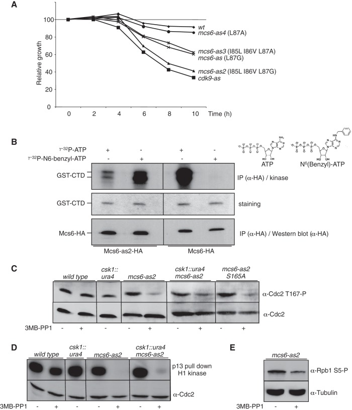
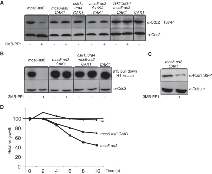
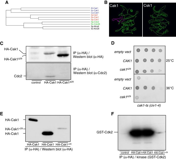
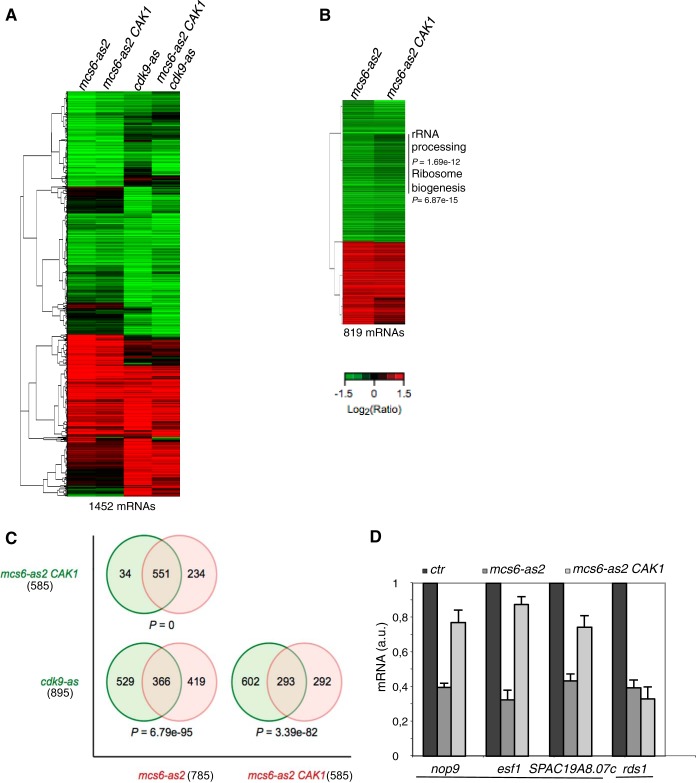
Cyclin-dependent kinase (Cdk) activation and RNA polymerase II transcription are linked by the Cdk7 kinase, which phosphorylates Cdks as a trimeric Cdk-activating kinase (CAK) complex, and serine 5 within the polymerase II (Pol II) C-terminal domain (CTD) as transcription factor TFIIH-bound CAK. However, the physiological importance of integrating these processes is not understood. Besides the Cdk7 ortholog Mcs6, fission yeast possesses a second CAK, Csk1. The two enzymes have been proposed to act redundantly to activate Cdc2. Using an improved analogue-sensitive Mcs6-as kinase, we show that Csk1 is not a relevant CAK for Cdc2. Further analyses revealed that Csk1 lacks a 20-amino-acid sequence required for its budding yeast counterpart, Cak1, to bind Cdc2. Transcriptome profiling of the Mcs6-as mutant in the presence or absence of the budding yeast Cak1 kinase, in order to uncouple the CTD kinase and CAK activities of Mcs6, revealed an unanticipated role of the CAK branch in the transcriptional control of the cluster of genes implicated in ribosome biogenesis and cell growth. The analysis of a Cdc2 CAK site mutant confirmed these data. Our data show that the Cdk7 kinase modulates transcription through its well-described RNA Pol II CTD kinase activity and also through the Cdc2-activating kinase activity.


Figures

FIG 1
FIG 1
Mcs6 is the genuine Cdc2-activating kinase in fission yeast. (A) Growth defect resulting from the inhibition of Mcs6-as. The indicated strains were grown in the presence or absence of 30 μM 3-MB-PP1. Cell numbers were measured and plotted relative to that of dimethyl sulfoxide (DMSO)-treated cells of the same genotype, defined as 100%. (B) In vitro kinase activity assay of the wt and analogue-sensitive (as) versions of Mcs6 in the presence of the wt or a bulky ATP analogue. HA-tagged versions of the Mcs6 or Mcs6-as2 kinases were precipitated (IP) using anti-HA antibodies and used on beads for kinase assays with either ATP or N6(benzyl)-ATP, with GST-CTD as the substrate. After separation on SDS-PAGE, the kinase gel was exposed (top), stained with Coomassie blue (middle), and transferred to a membrane for anti-HA (α-HA) Western blotting (bottom). Note that the exposure time of the left side of the kinase gel was shorter in order to compare the use of ATP versus N6(benzyl)-ATP. (C) In vivo effect of Mcs6 inactivation on Cdc2 T167 phosphorylation. The indicated strains were treated or not with the 3-MB-PP1 inhibitor, and total protein was extracted and separated on SDS-PAGE. Western blot analyses were performed with anti-Cdc2 T167P or anti-Cdc2, as indicated. (D) In vitro kinase assay of Cdc2 after inhibition of Mcs6. The indicated strains were treated or not with the 3-MB-PP1 inhibitor, and soluble proteins were extracted and immunoprecipitated using p13 beads. Kinase assays were performed using histone H1 as the substrate. After separation on SDS-PAGE, the kinase gel was exposed (top) and transferred to a membrane for anti-Cdc2 Western blotting (bottom). (E) Same as panel C, except that anti-S5P and anti tubulin were used in Western blotting.
FIG 2
FIG 2
Cak1 complements the CAK defect resulting from Mcs6 inactivation. (A) In vivo effects of Mcs6 inactivation on Cdc2 T167 phosphorylation are complemented by expression of Cak1. The indicated strains were grown in the presence or absence of 3-MB-PP1 inhibitor, and total protein was extracted and separated on SDS-PAGE. Western blot analyses were performed with anti-Cdc2 T167P or anti-Cdc2, as indicated. Note that the msc6-as2 data are identical to those in Fig. 1C and were duplicated here for clarity. All samples were run on the same gel. (B) In vitro kinase assay of Cdc2 after inhibition of Mcs6 in the presence of Cak1. The indicated strains were grown in the presence or absence of 3-MB-PP1 inhibitor, and soluble proteins were extracted and immunoprecipitated using p13 beads. Kinase assays were performed using histone H1 as the substrate. After separation on SDS-PAGE, the kinase gel was exposed (top) and transferred to a membrane for the anti-Cdc2 Western blotting (bottom). Note that the msc6-as2 data are identical to those in Fig. 1D and were duplicated here for clarity. All samples were run on the same gel. (C) The in vivo effect of Mcs6 inactivation on CTD phosphorylation is not complemented by expression of Cak1. Same as panel B, except that Western blot analysis was performed with anti-S5P or anti tubulin, as indicated. (D) The growth defect resulting from the inhibition of Mcs6-as is partially suppressed by Cak1. The indicated strains were grown in the presence or absence of 30 μM 3-MB-PP1. Cell numbers were measured and plotted relative to that of DMSO-treated cells of the same genotype, defined as 100%.
FIG 3
FIG 3
In vivo interaction between Cak1 and Cdc2 in fission yeast. (A) In vitro kinase activity assay of the wt and analogue-sensitive (as) versions of Cdk9 in the presence of wt or bulky ATP. TAP-tagged versions of the Cdk9 or Cdk9-as kinase were precipitated using anti-TAP antibodies and used on beads for kinase assays with either ATP or N6(benzyl)-ATP, with GST-CTD as the substrate. After separation on SDS-PAGE, the kinase gel was exposed. Note that the exposure time of the left side of the kinase gel was shorter in order to compare the use of ATP versus N6(benzyl)-ATP. (B) In vitro kinase activity assay of the wt and analogue-sensitive versions of Csk1 and Cak1 in the presence of wt or bulky ATP. HA-tagged versions of the indicated kinases were precipitated using anti-HA antibodies and used on beads for kinase assays with either ATP or N6(benzyl)-ATP, with GST-Cdk2 as the substrate. After separation on SDS-PAGE, the kinase gel was exposed (top and middle, with different exposure [exp] times) and transferred to a membrane for anti-HA Western blotting (bottom). (C) In vitro kinase activity assay of the wt versions of Csk1 and Cak1 in the presence of wt or bulky ATP. HA-tagged versions of the indicated kinases were precipitated using anti-HA antibodies and used on beads for kinase assays with either ATP or N6(benzyl)-ATP in the absence of exogenous substrate. After separation on SDS-PAGE, the kinase gel was exposed (middle) and transferred to a membrane for anti-HA Western blotting (top) or anti-Cdc2 Western blotting (bottom). The phosphorylated-substrate band was superimposed on the Cdc2 band.
FIG 4
FIG 4
A conserved insertion within the budding yeast Cak1 sequence family is required to bind Cdc2. (A) Phylogram tree based on a multiple alignment of the indicated protein sequences performed in ClustalW (not shown). Kl, K. lactis; Sc, S. cerevisiae; Ca, C. albicans; Sj, S. japonicus; So, S. octosporus; Sp, S. pombe. (B) Three-dimensional homology-based models of S. cerevisiae Cak1 (ScCak1) (left) and S. pombe Csk1 (SpCsk1) (right) built using SWISS-MODEL (http://swissmodel.expasy.org/?pid=smh01&uid=&token=). Only the backbone is represented. The conserved insertion present in Cak1 (see Fig. S3 in the supplemental material) is highlighted. The images were prepared with the Swiss-Pdb Viewer software. (C) Cdc2-binding assay of wt Cak1 and a truncated version lacking the sequence highlighted in panel B (Cak1Δ29). HA-tagged versions of the indicated kinases were precipitated using anti-HA antibodies. After SDS-PAGE and transfer to a membrane, they were processed for anti-HA Western blotting (top) and anti-Cdc2 Western blotting (bottom). (D) Complementation of a cak1 TS strain. The civ1-4 strain was transformed with pMET25 vectors expressing either CAK1 or cak1Δ29, and serial dilutions were plated at the indicated temperatures. (E) The Csk1+29 protein is expressed at a very low level. HA-tagged versions of the indicated kinases were precipitated using anti-HA antibodies. After SDS-PAGE and transfer to a membrane, they were processed for anti-HA Western blotting. (F) The Csk1+29 protein has low kinase activity. HA-tagged versions of the indicated kinases were precipitated using anti-HA antibodies and used on beads for kinase assays with GST-Cdk2 as the substrate. After separation on SDS-PAGE, the kinase gel was exposed.
FIG 5
FIG 5
Activation of Cdc2 by CAK is required for expression of the ribocluster. (A) Hierarchical clustering of 1,452 mRNAs whose expression is significantly affected in the mcs6-as2, mcs6-as2 CAK1, cdk9-as, and mcs6-as2 cdk9-as CAK1 mutant strains. The strains were cultured in yeast extract-supplemented medium, and the inhibitor 3-MB-PP1 or the solvent DMSO was added for 120 minutes. The data are presented as log2 30 μM 3-MB-PP1/DMSO ratios of hybridization intensity and are color coded as indicated in the key (a P value of ≤0.05 and ≥1.5-fold change). (B) Hierarchical clustering of the 819 mRNAs whose expression is significantly affected in the mcs6-as2 or the mcs6-as2 CAK1 strain (a P value of ≤0.05 and ≥1.5-fold change). The gene cluster whose expression is significantly decreased only in the absence of CAK1 is highlighted, and the enriched GO categories are indicated, with the associated P values. (C) Venn diagrams of the overlap in expression of mcs6-as2, mcs6-as2 CAK1, and cdk9-as with associated P values for the degree of overlap calculated by a hypergeometric test. The numbers of mRNAs are indicated in parentheses. (D) Relative quantification (RQ) of the nop9, esf1, SPAC19A8.07c, and rds1 mRNAs determined by quantitative RT-PCR using the ΔΔCT method in mcs6-as2 and mcs6-as2 CAK1 strains. Each strain was grown in the absence or presence of 3-MB-PP1 for 120 minutes, and treated samples were compared to untreated samples (ctr), set as 1. a.u., arbitrary units. Note that for clarity, the untreated control is shown only once per mRNA analyzed. The nop9 gene belongs to the rRNA-processing GO category (0006364), and the SPAC19A8.07c and esf1 genes belong to the ribosome biogenesis GO category (0042254). The error bars indicate standard deviations of the means from 3 independent experiments.
FIG 6
FIG 6
Activation of Cdc2 by CAK is required for the transcription of some ribosome-associated genes. (A) Dominant growth defect resulting from the expression of the cdc2 T167A mutant. (Top) A wt strain was transformed with plasmids expressing cdc2, cdc2 T167A (cdc2 TA), or empty vector, and serial dilutions were incubated at 32°C for 3 days. (Bottom) Colonies expressing the cdc2 TA mutant in the presence or absence of thiamine. Elongated cdc phenotypes (asterisk) appear only in the absence of thiamine. EMM, Edinburgh minimal medium. (B) RQ of the nop9, esf1, SPAC19A8.07c, and rds1 mRNAs determined by quantitative RT-PCR using the ΔΔCT method in the same strains as in panel A, grown in the presence of thiamine. The strain harboring the empty vector was set as 1. Note that for clarity, the untreated control is shown only once per mRNA analyzed. The nop9 gene belongs to the rRNA-processing GO category (0006364), and the SPAC19A8.07c and esf1 genes belong to the ribosome biogenesis GO category (0042254). The error bars indicate standard deviations of the means from 3 independent experiments.

Similar articles

Cited by

References

    1. Morgan DO. 1997. Cyclin-dependent kinases: engines, clocks, and microprocessors. Annu Rev Cell Dev Biol 13:261–291. doi:10.1146/annurev.cellbio.13.1.261. - DOI - PubMed
    1. Kaldis P. 1999. The cdk-activating kinase (CAK): from yeast to mammals. Cell Mol Life Sci 55:284–296. doi:10.1007/s000180050290. - DOI - PMC - PubMed
    1. Solomon MJ, Kaldis P. 1998. Regulation of CDKs by phosphorylation. Results Probl Cell Differ 22:79–109. doi:10.1007/978-3-540-69686-5_4. - DOI - PubMed
    1. Fisher RP. 2012. The CDK network: linking cycles of cell division and gene expression. Genes Cancer 3:731–738. doi:10.1177/1947601912473308. - DOI - PMC - PubMed
    1. Larochelle S, Batliner J, Gamble MJ, Barboza NM, Kraybill BC, Blethrow JD, Shokat KM, Fisher RP. 2006. Dichotomous but stringent substrate selection by the dual-function Cdk7 complex revealed by chemical genetics. Nat Struct Mol Biol 13:55–62. doi:10.1038/nsmb1028. - DOI - PubMed

Publication types

MeSH terms

Substances