. 2015 May;35(9):1573-87.
doi: 10.1128/MCB.00072-15. Epub 2015 Feb 17.

Requisite role for Nck adaptors in cardiovascular development, endothelial-to-mesenchymal transition, and directed cell migration

Affiliations

Requisite role for Nck adaptors in cardiovascular development, endothelial-to-mesenchymal transition, and directed cell migration

Derek L Clouthier et al. Mol Cell Biol. 2015 May.

Abstract

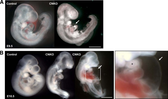
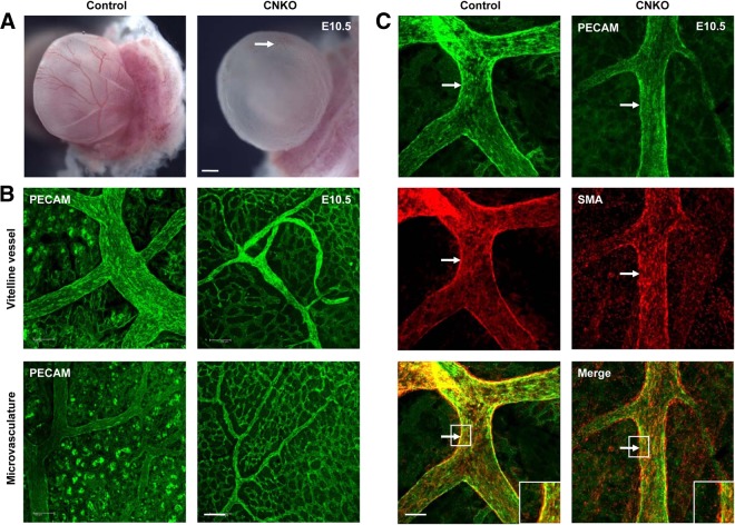
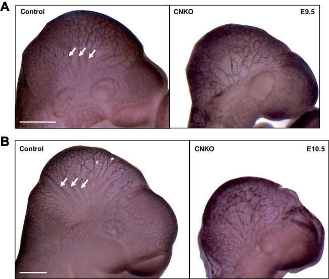
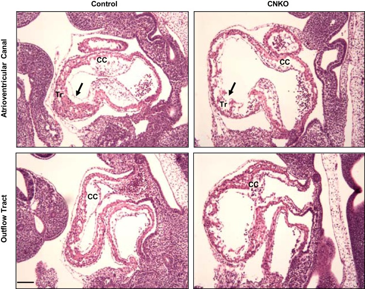
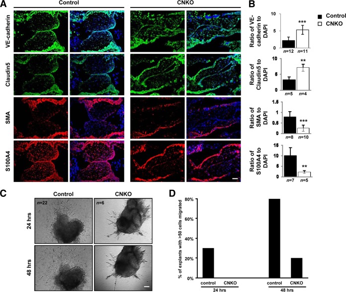
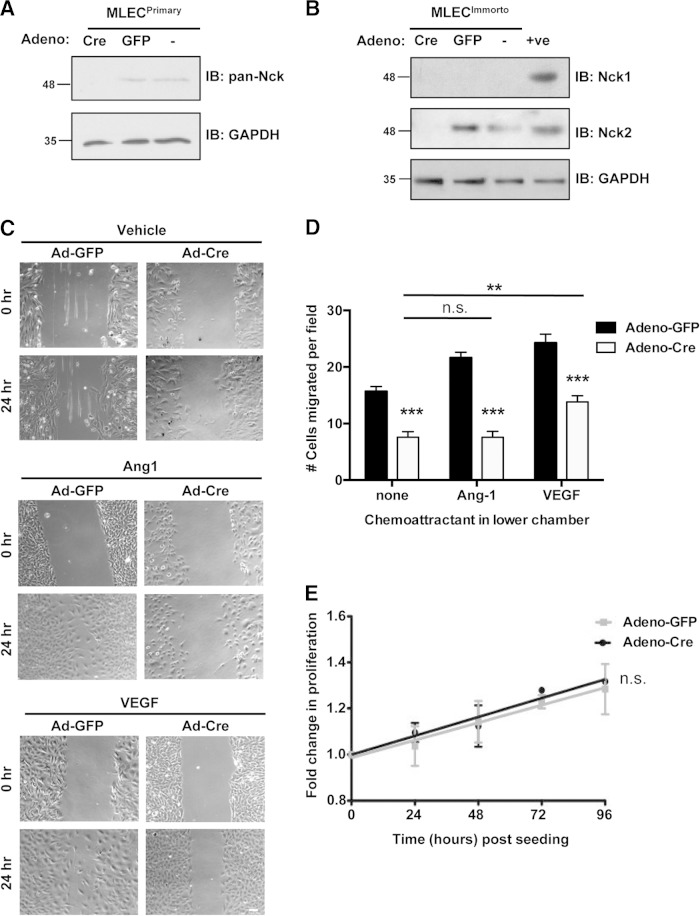
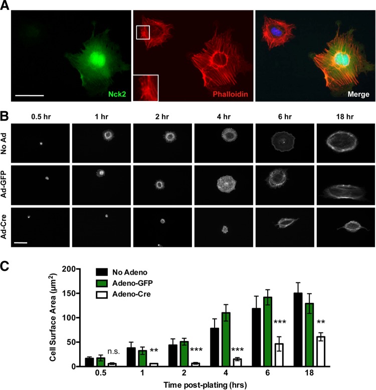
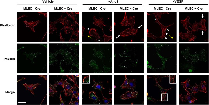
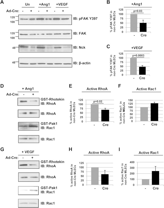
Development of the cardiovascular system is critically dependent on the ability of endothelial cells (ECs) to reorganize their intracellular actin architecture to facilitate migration, adhesion, and morphogenesis. Nck family cytoskeletal adaptors function as key mediators of actin dynamics in numerous cell types, though their role in EC biology remains largely unexplored. Here, we demonstrate an essential requirement for Nck within ECs. Mouse embryos lacking endothelial Nck1/2 expression develop extensive angiogenic defects that result in lethality at about embryonic day 10. Mutant embryos show immature vascular networks, with decreased vessel branching, aberrant perivascular cell recruitment, and reduced cardiac trabeculation. Strikingly, embryos deficient in endothelial Nck also fail to undergo the endothelial-to-mesenchymal transition (EnMT) required for cardiac valve morphogenesis, with loss of Nck disrupting expression of major EnMT markers, as well as suppressing mesenchymal outgrowth. Furthermore, we show that Nck-null ECs are unable to migrate downstream of vascular endothelial growth factor and angiopoietin-1, and they exhibit profound perturbations in cytoskeletal patterning, with disorganized cellular projections, impaired focal adhesion turnover, and disrupted actin-based signaling. Our collective findings thereby reveal a crucial role for Nck as a master regulator within the endothelium to control actin cytoskeleton organization, vascular network remodeling, and EnMT during cardiovascular development.


Figures

FIG 1
FIG 1
Generation and characterization of embryos lacking Nck expression in endothelial cells. (A) Breeding strategy used to generate conditional CNKO embryos with Nck deleted in the embryonic endothelium. The mutant genotype is Tie2-Cre+ Nck1−/− Nck2flox/flox (Nck2ff), and control genotypes are collectively Tie2-Cre Nck1+/ Nck2flox/flox, Tie2-Cre Nck1−/− Nck2flox/flox, and Tie2-Cre+ Nck1+/ Nck2flox/flox. (B) The efficiency of Cre-mediated Nck2 excision specifically within the vasculature of E10.5 CNKO embryos is shown via immunohistochemical analysis of Nck expression. Nck (green) is observed in the cardiac cushions (asterisks) and endocardial layer (arrows) of control but not CNKO embryos. (Insets) Images of the boxed areas highlighting the loss of Nck staining exclusively within the CNKO endocardium. Residual staining in CNKO embryos represents nonendothelial expression of Nck2. Cells are highlighted using DAPI (blue). (Bottom row) An adjacent section showing the endothelial marker claudin 5. Bar, 100 μm. (C) Proportion of viable versus nonviable CNKO embryos at E9.5, E10.5, and E11.5, as determined by the presence versus the absence of a heartbeat. The loss of endothelial Nck results in embryonic lethality between E10 and E11.
FIG 2
FIG 2
Conditional deletion of Nck in the endothelium results in embryonic lethality. Representative whole-mount images of control and CNKO embryos at E9.5 and E10.5 are shown. (A) At E9.5, CNKO embryos show underdevelopment of the heart, characterized by a reduced heart size, abnormal looping, and dilation of the pericardial cavity (arrow) compared to the findings for control embryos. (B) By E10.5, CNKO embryos are smaller than those of their control littermates, with hemorrhage and blood congestion being found throughout the body. The image on the right is an enlarged image of the boxed area highlighting severe pericardial edema (arrow) and disrupted cardiac chamber formation (asterisk). Bars, 1 mm.
FIG 3
FIG 3
CNKO yolk sacs exhibit decreased vessel remodeling and vessel integrity. (A) At E10.5, the yolk sacs of CNKO embryos appear pale in comparison to those of control embryos, owing to the underdevelopment of major and minor vessels (arrow). Bar, 1 mm. (B) Immunofluorescent PECAM staining of E10.5 yolk sacs demonstrates the reduced caliber of the vitelline vessel in CNKO yolk sacs compared to that of the controls, as well as poorly remodeled microvascular networks. Bar, 100 μm. (C) Dual immunofluorescence staining of E10.5 yolk sacs with PECAM and SMA shows an altered recruitment and association of smooth muscle cells with the vitelline vessel and major branches in CNKO yolk sacs compared to the findings for the controls (arrows and insets). Bar, 100 μm.
FIG 4
FIG 4
Defects in vascular remodeling in CNKO embryos. Whole-mount immunostaining of control and CNKO embryos for PECAM at E9.5 (20 to 25 somites) (A) or E10.5 (31 to 38 somites) (B). Control embryos show large-caliber major vessels in the head (arrows) at E9.5 with extensive branching (asterisks) by E10.5, while cranial vessels are underdeveloped and poorly organized in CNKO embryos. The images shown are representative of those for 14 embryos at E9.5 and 8 embryos at E10.5 for controls and of those for 5 embryos at E9.5 and 3 embryos at E10.5 for CNKO embryos. Bars, 500 μm (A) and 250 μm (B).
FIG 5
FIG 5
Characterization of cardiac defects in CNKO embryos. Hematoxylin-and-eosin-stained sagittal sections of control and CNKO embryos at E10.5 show the atrioventricular canal and outflow tract of the developing heart. CNKO embryos show a discontinuous endocardial lining (arrows), as well as reduced myocardial trabeculation (Tr), compared to the findings for the controls. Also, the well-defined cardiac cushions (CC) seen in the control embryos are hypoplastic in the CNKO embryos. Bar, 100 μm.
FIG 6
FIG 6
CNKO embryos show altered EnMT. (A) Immunofluorescent analysis of the endothelial markers VE-cadherin and claudin 5 (both green) and mesenchymal markers S100A4 and SMA (both red) within the AVC in serial sections obtained from E10.5 control and CNKO embryos, using DAPI (blue) counterstain. Bar, 50 μm. (B) Quantitative analysis of EnMT markers shows increased expression of VE-cadherin and claudin 5 and decreased expression of S100A4 and SMA in CNKO embryos compared to their levels of expression in the controls. The number of embryos analyzed is indicated below each histogram bar. **, P < 0.01; ***, P < 0.0001. (C) Phase-contrast images of AVC explants from E9.5 control or CNKO embryos at 24 and 48 h after plating on a collagen matrix. Bar, 100 μm. (D) Quantitative analysis of EnMT in AVC explants shows decreased cell outgrowth from CNKO explants compared to that for the controls at both time points. Individual explants were scored according to whether greater than or fewer than 50 cells had migrated into the collagen gels after plating.
FIG 7
FIG 7
Endothelial cells lacking Nck display defects in directed cell migration. (A) Immunoblots (IBs) of lysates obtained from primary MLECs transduced with Adeno-GFP or Adeno-Cre or left uninfected (−), showing a loss of Nck protein expression specifically within Adeno-Cre-infected cells (first lane). GAPDH shows equal protein loading of all MLEC samples. (B) Western blots of lysates obtained from conditionally immortalized MLECs transduced with Adeno-GFP or Adeno-Cre or left uninfected (−), showing that specific Cre-mediated Nck2 deletion results in the complete absence of Nck proteins (first lane). +ve, positive control. In panels A and B, numbers to the left of the blots are molecular masses (in kilodaltons). (C) Wound closure assay on confluent monolayers of primary MLECs transduced with either Adeno-GFP or Adeno-Cre and cultured on gelatin in the presence or absence of Ang1 or VEGF. Images of the same fields were taken at 0 and 24 h after wounding, and those shown are representative of the images from three independent experiments. Adeno-Cre-infected cells showed reduced wound closure under all conditions tested. Bar, 70 μm. (D) Boyden chamber assay on primary MLECs transduced with either Adeno-GFP or Adeno-Cre using either 100 ng/ml Ang1, 100 ng/ml VEGF, or no stimulus. The cells within at least seven random high-power fields were enumerated, and experiments were performed in triplicate. Adeno-Cre-infected cells showed a highly significant reduction (P < 0.001) in migration compared to Adeno-GFP-infected cells under all conditions tested, though a modest yet significant increase (P < 0.01) in migration of Adeno-Cre-infected cells could be seen with addition of VEGF compared to that seen with no chemoattractant. **, P < 0.01; ***, P < 0.0001; n.s., nonsignificant. (E) Proliferation assay on immortalized MLECs transduced with either Adeno-GFP or Adeno-Cre showing a nonsignificant difference in the growth rate in the presence or absence of Nck. Each time point represents data for 9 assays.
FIG 8
FIG 8
Altered actin organization and decreased cell spreading within Nck-deficient endothelial cells. (A) Representative fluorescence micrographs from a pool of Adeno-Cre-infected primary MLECs stained with anti-Nck2 antibodies (green), fluorescent phalloidin to show the morphology of F actin (red), and DAPI to highlight the position of the nucleus position (blue). Prominent parallel stress fibers and lamellipodial protrusions can be seen in Nck-expressing cells, while Nck-deficient MLECs are smaller, with stress fibers appearing short, disorganized, and highly bundled (inset). Bar, 30 μm. (B) Time course images of phalloidin-stained MLECs from panel A following 0.5, 1, 2, 4, 6, and 18 h of spreading on 0.1% gelatin-coated coverslips. Cells were simultaneously immunostained with Nck antibodies to confirm Nck2 excision. Adeno-Cre-infected MLECs show a reduced cell size with an altered actin morphology. Bar, 30 μm. (C) Quantitation of the results shown in panel B demonstrating that Adeno-Cre infection results in decreased cell spreading in comparison to that for Adeno-GFP-infected and uninfected (No Adeno) control cells. The histogram represents the mean cell surface area ± SEM for a minimum 6 to 10 cells per replicate. **, P < 0.01; ***, P < 0.0001; n.s., nonsignificant.
FIG 9
FIG 9
Aberrant focal adhesion remodeling in response to angiogenic stimuli in Nck-null MLECs. Representative fluorescence micrographs from immortalized MLECs that were transduced with Adeno-Cre (+Cre) or left uninfected (−Cre) and treated with 100 ng/ml Ang1 or 100 ng/ml VEGF for 15 min or vehicle alone. Fixed cells were stained with antipaxillin antibodies to highlight focal adhesion structures (green), fluorescent phalloidin to show the morphology of F actin (red), and DAPI to indicate the position of the nucleus (blue). Paxillin staining is found throughout the interior and periphery of unstimulated Nck-deficient MLECs, whereas it localizes exclusively to the periphery of control cells. Upon stimulation with Ang1 or VEGF, control cells assembled robust unidirectional lamellipodia (arrowheads) in the direction of cell movement (yellow arrows) which were accompanied by posterior stress fibers (asterisks), and focal adhesion structures were restricted to the tips of these projections (insets on merged images). In contrast, processes extended in multiple directions in Cre-infected cells (white arrows), and focal adhesions appeared larger, with paxillin punctae being localized around the entire cell periphery. Bar, 30 μm.
FIG 10
FIG 10
Endothelial Nck deficiency alters Rho GTPase and FAK activation. (A to C) Lysates obtained from immortalized MLECs (with or without Cre transduction) were treated with 100 ng/ml Ang1 or VEGF for 15 min and immunoblotted with antibodies recognizing pFAK Y397, FAK, Nck, or β-actin. Densitometric quantitation of the results in panel A demonstrates a significant decrease in FAK activation in Nck-null MLECs in response to Ang1 (B) and VEGF (C). β-Actin shows equal protein loading of all MLEC samples, and specific deletion of Nck upon Adeno-Cre transduction can also be observed. Results shown are representative of those from three to five independent experiments. In panel A, the numbers to the left of the blots are molecular masses (in kilodaltons). (D to I) Parallel lysates from panel A were incubated with GST-rhotekin or GST-Pak1 and immunoblotted with antibodies recognizing RhoA or Rac1. Densitometric quantitation of the results in panels D and G demonstrates a decrease in RhoA activation in Nck-null MLECs in response to Ang1 (E) and VEGF (H) and an increase in Rac1 activation in Nck-null MLECs in response to Ang1 (F) and VEGF (I). The relative amount of active Rac1 or RhoA in the Cre-transduced MLECs is represented as the percentage of the amount of active Rac1 or RhoA in control cells. Results are representative of those from three independent experiments.

References

    1. Carmeliet P. 2003. Blood vessels and nerves: common signals, pathways and diseases. Nat Rev Genet 4:710–720. doi:10.1038/nrg1158. - DOI - PubMed
    1. Carmeliet P. 2000. Mechanisms of angiogenesis and arteriogenesis. Nat Med 6:389–395. doi:10.1038/74651. - DOI - PubMed
    1. Armulik A, Abramsson A, Betsholtz C. 2005. Endothelial/pericyte interactions. Circ Res 97:512–523. doi:10.1161/01.RES.0000182903.16652.d7. - DOI - PubMed
    1. Srivastava D, Olson EN. 2000. A genetic blueprint for cardiac development. Nature 407:221–226. doi:10.1038/35025190. - DOI - PubMed
    1. Adams RH, Alitalo K. 2007. Molecular regulation of angiogenesis and lymphangiogenesis. Nat Rev Mol Cell Biol 8:464–478. doi:10.1038/nrm2183. - DOI - PubMed

Publication types

MeSH terms

LinkOut - more resources