. 2015 Mar 3;112(9):2800-5.
doi: 10.1073/pnas.1407163112. Epub 2015 Feb 17.

Ancient dynamin segments capture early stages of host-mitochondrial integration

Affiliations

Ancient dynamin segments capture early stages of host-mitochondrial integration

Ramya Purkanti et al. Proc Natl Acad Sci U S A. .

Abstract

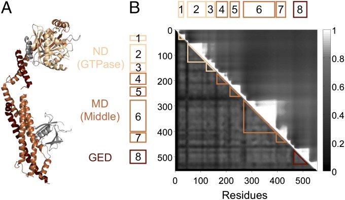
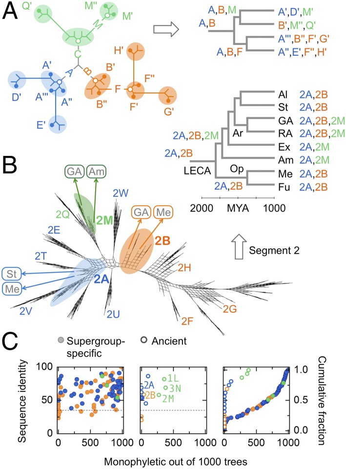
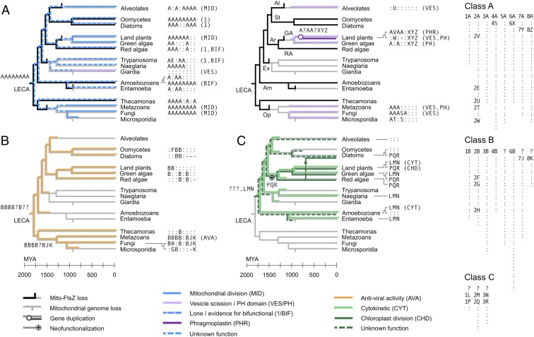
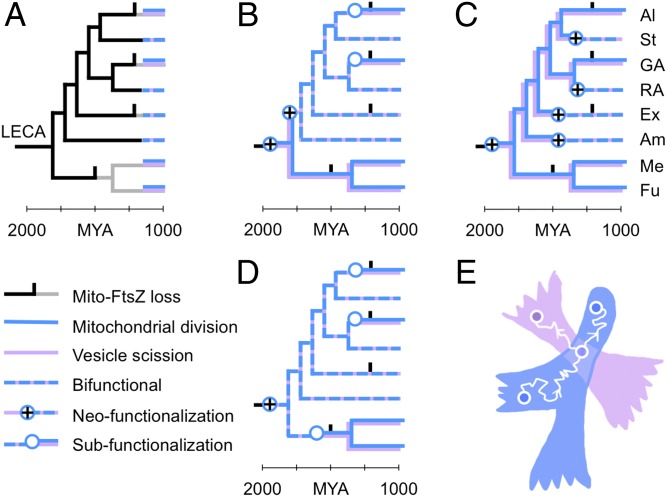
Eukaryotic cells use dynamins-mechano-chemical GTPases--to drive the division of endosymbiotic organelles. Here we probe early steps of mitochondrial and chloroplast endosymbiosis by tracing the evolution of dynamins. We develop a parsimony-based phylogenetic method for protein sequence reconstruction, with deep time resolution. Using this, we demonstrate that dynamins diversify through the punctuated transformation of sequence segments on the scale of secondary-structural elements. We find examples of segments that have remained essentially unchanged from the 1.8-billion-y-old last eukaryotic common ancestor to the present day. Stitching these together, we reconstruct three ancestral dynamins: The first is nearly identical to the ubiquitous mitochondrial division dynamins of extant eukaryotes, the second is partially preserved in the myxovirus-resistance--like dynamins of metazoans, and the third gives rise to the cytokinetic dynamins of amoebozoans and plants and to chloroplast division dynamins. The reconstructed sequences, combined with evolutionary models and published functional data, suggest that the ancestral mitochondrial division dynamin also mediated vesicle scission. This bifunctional protein duplicated into specialized mitochondrial and vesicle variants at least three independent times--in alveolates, green algae, and the ancestor of fungi and metazoans-accompanied by the loss of the ancient prokaryotic mitochondrial division protein FtsZ. Remarkably, many extant species that retain FtsZ also retain the predicted ancestral bifunctional dynamin. The mitochondrial division apparatus of such organisms, including amoebozoans, red algae, and stramenopiles, seems preserved in a near-primordial form.

Keywords: FtsZ; dynamin; eukaryote evolution; mitochondria; vesicles.


Conflict of interest statement

The authors declare no conflict of interest.

Figures

Fig. 1.
Fig. 1.
Breaking dynamin into evolutionary segments. (A) The crystal structure of human dynamin 1 [Protein Data Bank (PDB) ID: 3SNH] (18). Dynamins have three conserved domains: the N-terminal GTPase domain (ND, light brown), the middle domain (MD, brown), and the GTPase effector domain (GED, dark brown). (B) We assemble a 556-residue concatenated alignment of the ND, MD, and GED. Our analysis relies on finding clusters of proteins with high sequence identity across short segments of this alignment. Two contiguous segments with similar clusters can be merged into a single longer segment with no loss of information. The upper-triangular matrix Rij shows the properties of long segments starting at residue i and ending at residue j; high values indicate that the long segment is made up of multiple subsegments with similar clustering properties (SI Text, Evolutionary Segments). The saw tooth structure shows that dynamin breaks into eight natural evolutionary segments ranging from 22 to 118 residues in length: three in the ND, four in the MD, and one in the GED. Every dynamin is assigned an eight-letter signature, such that two proteins with the same letter at a given segment have high sequence identity across that segment (SI Text, Sequence Clustering).
Fig. 2.
Fig. 2.
Living fossils: tight, ancient, paraphyletic clusters. (A) (Left) Schematic protein tree. Proteins are grouped into clusters of high pairwise identity corresponding to letter labels (colored ovals and circles). For each cluster we show extant proteins (solid dots) and their last common ancestor (root proteins: open dots). If descendants of a root protein are confined to the cluster itself, the cluster is monophyletic; otherwise it is paraphyletic, with other clusters branching out of it. (Right) Extant proteins are mapped to the leaves of a species tree. If a cluster contains proteins from multiple species, their last common ancestor is its root species. Three superclasses (blue, orange, and green) emerge from three root proteins (“A,” “B,” and “C”). Proteins similar to A and B survive to the present day, and no proteins similar to C survive. (B) Maximum-likelihood trees for one trial of segment 2. We show a consensus split network (48) representing 100 bootstrap replicates; nodes with strong bootstrap support appear as thin stems. Ancient clusters (2A, 2B, and 2M) are shown with colored overlays and the rest with letters; not all clusters are labeled. Paraphyletic clusters 2A and 2B form “galls” from which other class A (blue) and class B (orange) clusters emerge; monophyletic clusters like 2M have no outward branches. Clusters 2U and 2W map to the center of the tree due to long-branch attraction (Fig. S3D). We map ancestral proteins to a species tree (Mya: millions of years ago); species group labels are as in Fig. 3. Ancient clusters contain proteins from multiple supergroups (labels in boxes). (C) To find the phylogenetic attributes of clusters corresponding to each letter label, the analysis shown in Fig. 2B for segment 2 is repeated for each segment separately. We show the attributes of all 110 clusters in our dataset with more than 10 members. Attribute 1: pairwise identity (y axis, Left and Center; dashed line, 35% identity). Attribute 2: root species (open circles, ancient, root species >1.5 billion y old; solid circles, supergroup-specific, root species <1.5 billion y old). Attribute 3: monophyly (x axis, Left, Center, and Right, monophyly support out of 1,000 trees; Right, cumulative distribution of monophyly support). If a cluster is tight (pairwise identity >35%), ancient (root species >1.5 billion y old), and paraphyletic (monophyly support <∼100/1,000), its members are living fossils: extant proteins similar in sequence to the ancient root proteins of functional superclasses. All ancient class A and class B clusters (e.g., 2A and 2B, Center) are paraphyletic and therefore ancestral. The ancient class C clusters (1L, 2M, and 3N) are monophyletic.
Fig. 3.
Fig. 3.
Punctuated diversification of dynamins across 1.8 billion y. The heterogeneous nature of dynamin evolution is highlighted, over time and across the protein sequence. The evolutions of different dynamin superclasses (colored) and of mitochondrial FtsZ (black) are overlaid on the eukaryotic tree (gray). The timing of speciation events, from 2,000 million years ago (Mya) to the present day, is approximately as reported in Parfrey et al. (11); the FtsZ loss data are derived from Fig. S6. Certain events (mitochondrial genome loss, FtsZ loss, other gene loss and duplication events, and horizontal transfers) are shown on the correct branches but not necessarily at the correct times. Species group labels: Al, alveolates; St, stramenopiles; Ar, archaeplastids; GA, green algae (including land plants); RA, red algae; Ex, excavates; Am, amoebozoans; Op, opisthokonts; Me, metazoans; Fu, fungi. We represent dynamins with 8-letter signatures (Dataset S1, sheets 1 and 2; class C dynamins span only segments 1–3). Each segment-wise letter labels a cluster of similar sequences; two proteins with the same letter at a given segment thus have high sequence identity across that segment. The key (Right) shows the full set of 238 letter clusters across segments and superclasses, sorted as in Dataset S1, sheet 2. The letters A and B represent distinct clusters at each segment, corresponding to the reconstructed root proteins of classes A and B; the root protein of class C cannot be reconstructed. Most supergroup-specific clusters are labeled with the shorthand symbol “:” but are assigned full cluster labels in Dataset S1; no two instances of : in this figure correspond to the same full label. A subset of clusters, including all those spanning multiple supergroups, is labeled by segment-wise letters for ease of reference. Gaps are labeled “-”; segments of ancestral proteins that cannot be reconstructed are labeled “?.” We show functional annotations [mitochondrial division (MID), vesicle scission (VES), etc.] where they have been experimentally verified (Table S1). (A) (Left) Class A dynamins specialized for mitochondrial division (MID; solid blue), or lone/bifunctional (1/BIF; dashed blue and lavender; “lone” indicates a single copy per genome). (Right) Derived class A dynamins specialized for vesicle scission (VES; lavender) or phragmoplast/cell plate formation [phragmoplastin (PHR); purple]. (B) Class B dynamins involved in antiviral activity (AVA) (orange). (C) Class C dynamins specialized for cytokinesis (CYT) (green) or chloroplast division (CHD) (dark green). The cytokinetic (LMN) and plastid division dynamins (PQR) are sister groups, descended from an ancestral dynamin of unknown sequence (???) (Fig. S2). The vertical arrow shows horizontal gene transfer by secondary endosymbiosis of red algae by diatoms (Bacillariophyta). Some rootings of the eukaryotic tree would place the ancestral class C dynamin at LECA itself (Fig. S4).
Fig. 4.
Fig. 4.
Duplication and specialization of a bifunctional ancestral dynamin. (A) Present-day lineages have either a bifunctional dynamin with both mitochondrial and vesicle roles (dashed blue and lavender) or two dynamins specialized for mitochondrial division and vesicle scission (stacked blue and lavender). We want to find the most parsimonious explanation for this distribution. Scenarios using alternative rootings of the eukaryotic tree are shown in Fig. S4. Species group labels are as in Fig. 3. (B and C) Scenarios in which LECA had two specialized dynamins require multiple gain-of-function events. (D) The scenario in which LECA had a single bifunctional dynamin predicts that specialized dynamins emerge from at least three independent subfunctionalization events, all coupled with the loss of FtsZ. The causal relationship between gene duplication and FtsZ loss is not clear. (E) A 2D schematic of protein sequence space. A single protein sequence is represented as a point (circles); as its evolves by random mutation, the corresponding point traces a curve through the space (white wiggly arrows represent the passage of time). Some amino acid sequences do not encode functional proteins (white background); other regions encode mitochondrial dynamins (X; blue) and vesicle dynamins (Y; lavender). If such regions overlap, it implies the possibility of a bifunctional dynamin. A protein starts in one of the monofunctional regions (bottom left circle). As it diffuses through sequence space (white wiggly arrow) it is confined to this region by purifying selection for function X. Eventually, it discovers a bifunctional region XY, in the overlap of the blue and lavender regions (middle circle). If the protein then duplicates (branch), the two new copies subfunctionalize by rapidly exiting the bifunctional region, back into the blue and lavender regions (white wiggly arrows moving toward the top left and right circles; each arrow represents a distinct protein copy). The dynamics of this process are derived in SI Text, Routes of Gene Duplication.

References

    1. Williams TA, Foster PG, Cox CJ, Embley TM. An archaeal origin of eukaryotes supports only two primary domains of life. Nature. 2013;504(7479):231–236. - PubMed
    1. Baum DA, Baum B. An inside-out origin for the eukaryotic cell. BMC Biol. 2014;12:76. - PMC - PubMed
    1. Ramadas R, Thattai M. New organelles by gene duplication in a biophysical model of eukaryote endomembrane evolution. Biophys J. 2013;104(11):2553–2563. - PMC - PubMed
    1. Keeling PJ. Diversity and evolutionary history of plastids and their hosts. Am J Bot. 2004;91(10):1481–1493. - PubMed
    1. Osteryoung KW, Nunnari J. The division of endosymbiotic organelles. Science. 2003;302(5651):1698–1704. - PubMed

Publication types