. 2015:2015:460610.
doi: 10.1155/2015/460610. Epub 2015 Jan 27.

Deficits in endogenous adenosine formation by ecto-5'-nucleotidase/CD73 impair neuromuscular transmission and immune competence in experimental autoimmune myasthenia gravis

Affiliations

Deficits in endogenous adenosine formation by ecto-5'-nucleotidase/CD73 impair neuromuscular transmission and immune competence in experimental autoimmune myasthenia gravis

Laura Oliveira et al. Mediators Inflamm. 2015.

Abstract

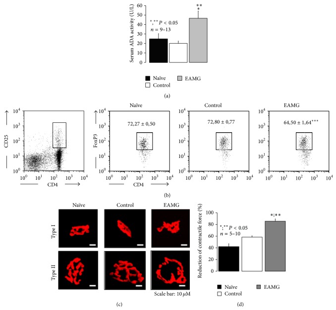
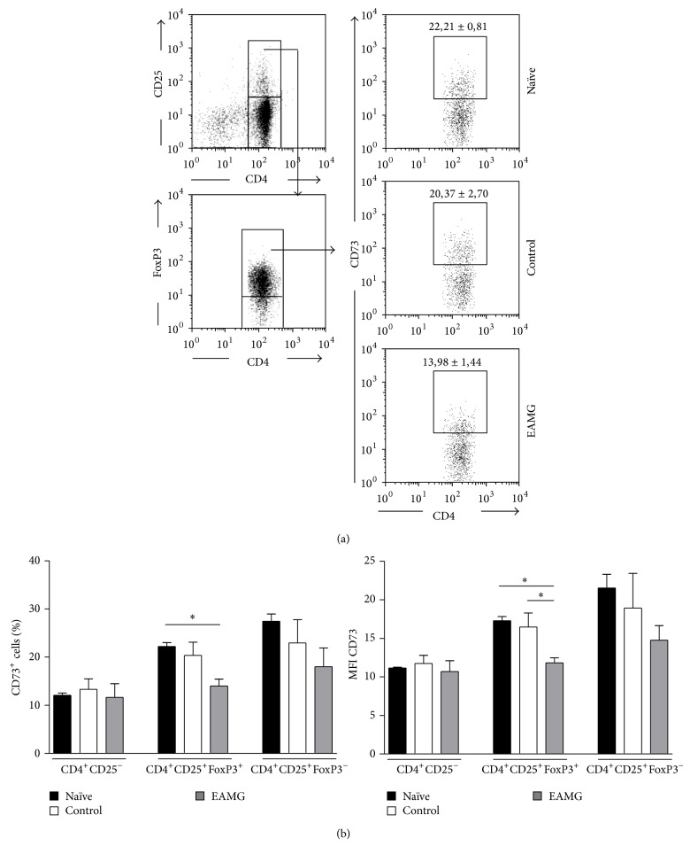
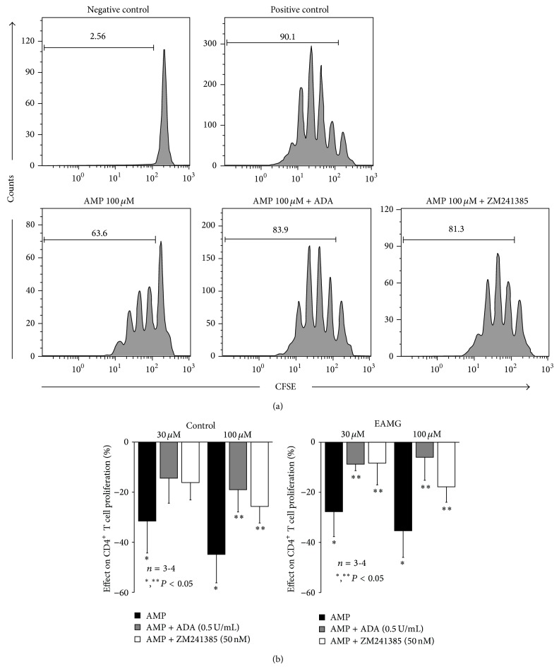
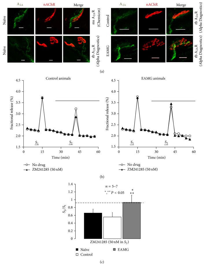
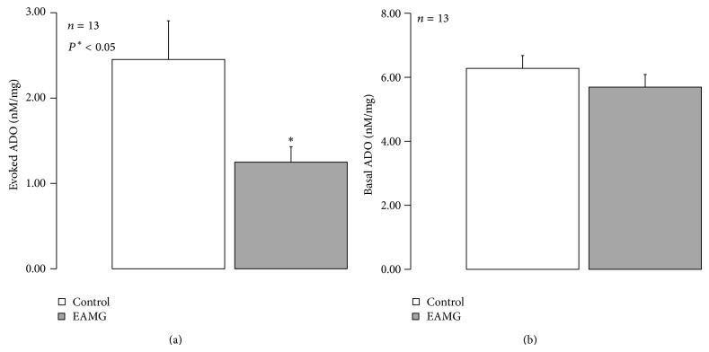
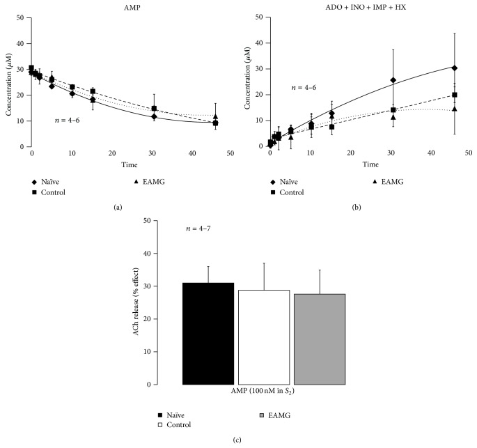
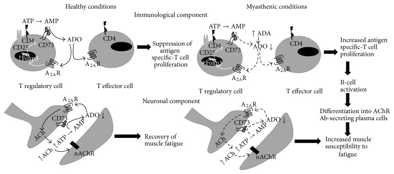
AMP dephosphorylation via ecto-5'-nucleotidase/CD73 is the rate limiting step to generate extracellular adenosine (ADO) from released adenine nucleotides. ADO, via A2A receptors (A2ARs), is a potent modulator of neuromuscular and immunological responses. The pivotal role of ecto-5'-nucleotidase/CD73, in controlling extracellular ADO formation, prompted us to investigate its role in a rat model of experimental autoimmune myasthenia gravis (EAMG). Results show that CD4(+)CD25(+)FoxP3(+) regulatory T cells express lower amounts of ecto-5'-nucleotidase/CD73 as compared to controls. Reduction of endogenous ADO formation might explain why proliferation of CD4(+) T cells failed upon blocking A2A receptors activation with ZM241385 or adenosine deaminase in EAMG animals. Deficits in ADO also contribute to neuromuscular transmission failure in EAMG rats. Rehabilitation of A2AR-mediated immune suppression and facilitation of transmitter release were observed by incubating the cells with the nucleoside precursor, AMP. These findings, together with the characteristic increase in serum adenosine deaminase activity of MG patients, strengthen our hypothesis that the adenosinergic pathway may be dysfunctional in EAMG. Given that endogenous ADO formation is balanced by ecto-5'-nucleotidase/CD73 activity and that A2ARs exert a dual role to restore use-dependent neurocompetence and immune suppression in myasthenics, we hypothesize that stimulation of the two mechanisms may have therapeutic potential in MG.


Figures

Figure 1
Figure 1
Neuromuscular and immunological deficits of rats with EAMG. Experiments were performed six weeks after immunization with the peptide R97-116 corresponding to the α-subunit of nAChR in CFA (EAMG), as compared to age-matched naïve and control littermates. (a) Measurement of serum adenosine deaminase (ADA) activity. Results are mean ± SEM of 9 naïve, 13 control, and 12 EAMG rats. *, ** P < 0.05 (one-way ANOVA following Dunnett's modified t-test) compared to naïve and control animals, respectively. (b) Flow cytometry analysis of intracellular FoxP3 expression in CD4+CD25+  T cells collected from popliteal and inguinal lymph nodes from naïve, control, and EAMG rats. Gating strategy to delimit CD4+CD25+ cells is shown on the left dot plot. Dot plots on the right show FoxP3 expression within gated CD4+CD25+ cells. Gate inside these dot plots correspond to FoxP3+ cells. Dot plots are a representative example of each indicated group. Numbers inside dot plots correspond to mean percentage ± SEM of FoxP3+ cells within CD4+CD25+ population. Statistically significant difference between EAMG (n = 13) and both naïve (n = 9) and control (n = 13) groups is indicated (*** P < 0.001; one-way ANOVA and Bonferroni's post hoc test). (c) Confocal microscopy micrographs showing type I and type II motor endplates from hemidiaphragm sections labeled with TMR-α-BTX (red) from naïve, control, and EAMG animals. Scale bar: 10 μm. (d) Reduction of contractile strength of isolated hemidiaphragm preparations from naïve, control, and EAMG animals during a 3 min period of intermittent phrenic nerve stimulation (17 pulses per second delivered at 50 Hz frequency). Results are mean ± SEM of 10 naïve, 6 control, and 5 EAMG rats. *, ** P < 0.05 (one-way ANOVA following Dunnett's modified t-test) compared to naïve and control animals, respectively.
Figure 2
Figure 2
(a) Flow cytometry analysis of surface CD73 expression on CD4+CD25+FoxP3+  T cells of popliteal and inguinal lymph nodes from naïve, control, and EAMG animals. Gating strategies are indicated on dot plots on the left. Dot plots on the right show CD73 expression within CD4+CD25+FoxP3+  T cells and are a representative example of each indicated group. Numbers inside dot plots correspond to mean percentage ± SEM of CD73+ cells. (b) Percentage of CD73+ cells (left) and mean fluorescence intensity (MFI) due to CD73 (right) staining within the indicated cell populations. Bars represent means ± SEM of 5 experiments for each animal group. * P < 0.05, ** P < 0.01 (one-way ANOVA and Bonferroni's post hoc test) compared to naïve and control animals, respectively.
Figure 3
Figure 3
(a) Flow cytometric evaluation of plated anti-CD3 and soluble anti-CD28 mAbs (1 μg/mL) induced proliferative response of 5 × 10⁴ CFSE-labelled CD4+T cells, sorted from popliteal and inguinal lymph nodes of a control animal, cultured for 3 days in the absence (positive control) or presence of AMP (100 μM), AMP (100 μM) plus ADA (0,5 U/mL), or AMP (100 μM) plus ZM241385 (50 nM), as indicated. Negative control corresponds to unstimulated cells (no mAbs added). Numbers within histograms correspond to the percentage of cells that divided at least once. Results shown are a representative example of 3 to 4 independent experiments performed in different animals. (b) AMP induced inhibition of CD4+ T cell proliferation obtained from popliteal and inguinal lymph nodes from control and EAMG animals. Bars represent means ± SEM of 3 control and 4 EAMG animals. * P < 0.05 compared to the absence of AMP and ** P < 0.05 compared to the AMP effect (one-way ANOVA and Bonferroni's post hoc test).
Figure 4
Figure 4
Tonic activation of A2AR is significantly impaired at motor endplates of myasthenic rats. (a) Confocal micrographs showing immunoreactivity against A2AR (green) on motor endplates from rat hemidiaphragms labeled with TMR-α-BTX (red) from naïve, control, and EAMG rats. Scale bar: 10 μm. Two distinct A2AR antibodies, AlphaDiagnostics (A2aR21-P) and Chemicon (05-717, Clone 7F6-G5-A2), were used was indicated. (b) Time course of tritium outflow from phrenic nerve terminals from control and EAMG animals taken from typical experiments in the absence (no drug, open circles) and in the presence of the selective A2AR antagonist, ZM241285 (50 nM) (filled triangles). [3H]-ACh release was elicited by stimulating the phrenic nerve trunk with 750 pulses delivered with a frequency of 5 Hz at the indicated times (S 1 and S 2). ZM241285 (50 nM) was applied 15 min before S 2. (c) Modification of the S 2/S 1 ratio caused by ZM241285 (50 nM) in naïve, control, and EAMG rats. Each column represents pooled data from five (naïve and control) and seven (EAMG) animals. The vertical bars represent mean ± SEM. *, ** P < 0.05 (one-way ANOVA followed by Dunnett's modified t-test) when compared to naïve and control (CFA) rats.
Figure 5
Figure 5
The amount ADO released upon phrenic nerve stimulation is lower in EAMG animals. The ordinates represent the (a) evoked release of ADO upon phrenic nerve trunk electrical stimulation (750 pulses applied at 5 Hz frequency) and (b) basal ADO quantified by HPLC(diode array detection). Nerve-evoked release of ADO was calculated by subtracting the basal release, measured in the sample collected before stimulation, from the total release of adenosine determined after stimulus application. The data are means ± S.E.M. of 13 animals of each group (naïve, control, and EAMG).
Figure 6
Figure 6
Dephosphorylation of AMP, to ADO via ecto-5′-nucleotidase/CD73, facilitates the release of [3H]-ACh from stimulated phrenic motor nerve terminals of naïve, control, and EAMG rats. (a) and (b) show the kinetics of the extracellular AMP (30 μM) catabolism (a) and formation of ADO plus nucleoside derivatives (inosine (INO) and hypoxanthine (HX)) (b) in hemidiaphragm preparations from naïve, control, and EAMG rats. AMP (30 μM) was added to the preparation at zero time; samples were collected from the bath at indicated times on the abscissa and retained for HPLC analysis. Data shown are averages pooled from 4 naïve, 6 control, and 6 EAMG animals. The vertical bars represent SEM and are shown when they exceed the symbols in size. (c) Facilitatory effects of AMP (100 μM) on evoked [3H]-ACh release (5 Hz, 750 pulses, S 1 and S 2) from hemidiaphragm preparations from naïve, control, and EAMG rats. AMP (100 μM) was applied 15 min before S 2. Each column represents pooled data from 4 (naïve and control) and 7 (EAMG) animals. The vertical bars represent mean ± SEM.
Figure 7
Figure 7
Participation of the adenosinergic system on neuroimmunological deficits present in EAMG rats. In healthy animals, A2AR activation by ADO generated from the catabolism of released nucleotides via ecto-5′-nucleotidase/CD73 downmodulates T effector (CD4+CD25+FoxP3) cells proliferation in response to specific antigens by increasing the activity of Treg (CD4+CD25+FoxP3+) cells expressing FoxP3-dependent gene products, like ecto-5′-nucleotidase/CD73. At the neuromuscular junction, ecto-5′-nucleotidase/CD73 activity leads to the formation of ADO from released ATP (from both nerve and muscle), which facilitates acetylcholine release via prejunctional A2AR activation that is necessary to resist tetanic depression. In EAMG rats, increases in serum adenosine deaminase (ADA) together with ecto-5′-nucleotidase/CD73 in Treg (CD4+CD25+FoxP3+) cells lead to insufficient amounts of extracellular ADO. The lack of the A2AR immunosuppressive tonus contributes to the loss of peripheral tolerance to nAChR. Thus, increases in the proliferation of antigen-specific T effector cells triggers B cells differentiation into plasma cells and secretion of antibodies directed towards motor endplates nAChR clusters. This antibody attack leads to nAChR internalization/degradation and to complement-mediated morphological changes of the myasthenic postsynaptic membrane (e.g., fewer secondary synaptic folds, widening of the synaptic cleft). These changes contribute to neuromuscular transmission failure, which is further aggravated by deficits in the production of extracellular ADO, probably from released adenine nucleotides, namely, ATP. Impairment of tonic A2AR-mediated facilitation of transmitter Impairment of tonic A2AR-mediated facilitation of transmitter release turns myasthenic skeletal muscles unable to resist fatigue.

Similar articles

Cited by

References

    1. Lindstrom J. M. Acetylcholine receptors and myasthenia. Muscle & Nerve. 2000;23(4):453–477. - PubMed
    1. Conti-Fine B. M., Milani M., Kaminski H. J. Myasthenia gravis: past, present, and future. Journal of Clinical Investigation. 2006;116(11):2843–2854. doi: 10.1172/JCI29894. - DOI - PMC - PubMed
    1. Hirsch N. P. Neuromuscular junction in health and disease. British Journal of Anaesthesia. 2007;99(1):132–138. doi: 10.1093/bja/aem144. - DOI - PubMed
    1. Correia-de-Sá P., Sebastião A. M., Ribeiro J. A. Inhibitory and excitatory effects of adenosine receptor agonists on evoked transmitter release from phrenic nerve endings of the rat. British Journal of Pharmacology. 1991;103(2):1614–1620. doi: 10.1111/j.1476-5381.1991.tb09836.x. - DOI - PMC - PubMed
    1. Csóka B., Himer L., Selmeczy Z., et al. Adenosine A2A receptor activation inhibits T helper 1 and T helper 2 cell development and effector function. The FASEB Journal. 2008;22(10):3491–3499. doi: 10.1096/fj.08-107458. - DOI - PMC - PubMed

Publication types