Computed tomography enterography and magnetic resonance enterography in the diagnosis of Crohn's disease
- PMID: 25691841
- PMCID: PMC4316219
- DOI: 10.5217/ir.2015.13.1.27
Computed tomography enterography and magnetic resonance enterography in the diagnosis of Crohn's disease
Abstract
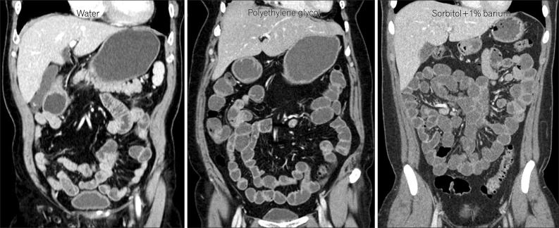
Imaging of the small bowel is complicated by its length and its overlapping loops. Recently, however, the development of crosssectional imaging techniques, such as computed tomography enterography (CTE) and magnetic resonance enterography (MRE) has shifted fundamental paradigms in the diagnosis and management of patients with suspected or known Crohn's disease (CD). CTE and MRE are noninvasive imaging tests that involve the use of intraluminal oral and intravenous contrast agents to evaluate the small bowel. Here, we review recent advances in each cross-sectional imaging modality, their advantages and disadvantages, and their diagnostic performances in the evaluation of small bowel lesions in CD.
Keywords: Crohn disease; Inflammatory bowel diseases; Magnetic resonance imaging; Tomography, spiral computed.
Conflict of interest statement
Conflict of interest: None.
Figures

Similar articles
-
Interobserver and intermodality agreement for detection of small bowel Crohn's disease with MR enterography and CT enterography.Inflamm Bowel Dis. 2011 May;17(5):1081-8. doi: 10.1002/ibd.21534. Epub 2010 Nov 15. Inflamm Bowel Dis. 2011. PMID: 21484959 Clinical Trial.
-
Role of computed tomography enterography/magnetic resonance enterography: is it in prime time?Clin Endosc. 2012 Sep;45(3):269-73. doi: 10.5946/ce.2012.45.3.269. Epub 2012 Aug 22. Clin Endosc. 2012. PMID: 22977815 Free PMC article.
-
Is the Mixed Use of Magnetic Resonance Enterography and Computed Tomography Enterography Adequate for Routine Periodic Follow-Up of Bowel Inflammation in Patients with Crohn's Disease?Korean J Radiol. 2022 Jan;23(1):30-41. doi: 10.3348/kjr.2021.0072. Epub 2021 Sep 13. Korean J Radiol. 2022. PMID: 34564963 Free PMC article.
-
Magnetic resonance enterography for the evaluation of the deep small intestine in Crohn's disease.Intest Res. 2016 Apr;14(2):120-6. doi: 10.5217/ir.2016.14.2.120. Epub 2016 Apr 27. Intest Res. 2016. PMID: 27175112 Free PMC article. Review.
-
Computed Tomography and Magnetic Resonance Small Bowel Enterography: Current Status and Future Trends Focusing on Crohn's Disease.Gastroenterol Clin North Am. 2018 Sep;47(3):475-499. doi: 10.1016/j.gtc.2018.04.002. Epub 2018 Jul 7. Gastroenterol Clin North Am. 2018. PMID: 30115433 Review.
Cited by
-
The Diagnostic Role of Magnetic Resonance Enterography as a Complementary Test to Colonoscopy in Active Crohn's Disease.Middle East J Dig Dis. 2016 Apr;8(2):93-101. doi: 10.15171/mejdd.2016.13. Middle East J Dig Dis. 2016. PMID: 27252815 Free PMC article.
-
Low-Dose Computed Tomography for the Optimization of Radiation Dose Exposure in Patients with Crohn's Disease.Gastroenterol Res Pract. 2018 Oct 31;2018:1768716. doi: 10.1155/2018/1768716. eCollection 2018. Gastroenterol Res Pract. 2018. PMID: 30515203 Free PMC article. Review.
-
Computed Tomography Enterography: Quantitative Evaluation on Crohn's Disease Activity.Gastroenterol Res Pract. 2018 Jul 22;2018:7351936. doi: 10.1155/2018/7351936. eCollection 2018. Gastroenterol Res Pract. 2018. PMID: 30140280 Free PMC article.
-
Assessment of Disease Activity in Small Bowel Crohn's Disease: Comparison between Endoscopy and Magnetic Resonance Enterography Using MRIA and Modified MRIA Score.Gastroenterol Res Pract. 2015;2015:159641. doi: 10.1155/2015/159641. Epub 2015 Nov 22. Gastroenterol Res Pract. 2015. PMID: 26759554 Free PMC article.
-
Evaluating inflammatory activity in Crohn's disease by cross-sectional imaging techniques.Radiol Bras. 2020 Jan-Feb;53(1):38-46. doi: 10.1590/0100-3984.2018.0096. Radiol Bras. 2020. PMID: 32313336 Free PMC article.
References
-
- Raptopoulos V, Schwartz RK, McNicholas MM, Movson J, Pearlman J, Joffe N. Multiplanar helical CT enterography in patients with Crohn's disease. AJR Am J Roentgenol. 1997;169:1545–1550. - PubMed
-
- Paulsen SR, Huprich JE, Fletcher JG, et al. CT enterography as a diagnostic tool in evaluating small bowel disorders: review of clinical experience with over 700 cases. Radiographics. 2006;26:641–657. - PubMed
-
- Maglinte DD, Sandrasegaran K, Lappas JC. CT enteroclysis: techniques and applications. Radiol Clin North Am. 2007;45:289–301. - PubMed
-
- Wold PB, Fletcher JG, Johnson CD, Sandborn WJ. Assessment of small bowel Crohn disease: noninvasive peroral CT enterography compared with other imaging methods and endoscopy--feasibility study. Radiology. 2003;229:275–281. - PubMed
-
- Negaard A, Sandvik L, Berstad AE, et al. MRI of the small bowel with oral contrast or nasojejunal intubation in Crohn's disease: randomized comparison of patient acceptance. Scand J Gastroenterol. 2008;43:44–51. - PubMed
Publication types
LinkOut - more resources
Full Text Sources
Other Literature Sources