

Neurogenic niches in the brain: help and hindrance of the barrier systems
- PMID: 25691856
- PMCID: PMC4315025
- DOI: 10.3389/fnins.2015.00020
Neurogenic niches in the brain: help and hindrance of the barrier systems
Abstract
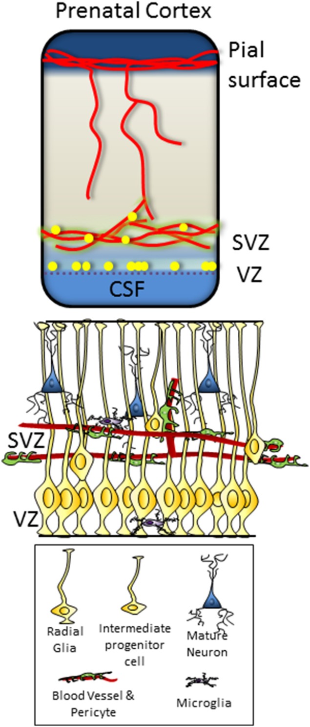
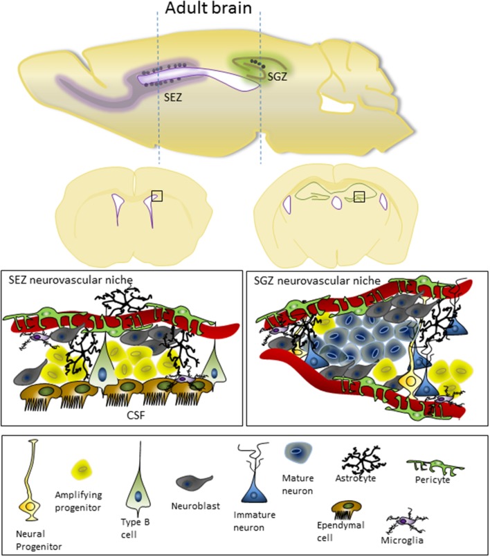
In the developing central nervous system, most neurogenesis occurs in the ventricular and subventricular proliferative zones. In the adult telencephalon, neurogenesis contracts to the subependyma zone and the dentate gyrus (subgranular zone) of the hippocampus. These restricted niches containing progenitor cells which divide to produce neurons or glia, depending on the intrinsic and environmental cues. Neurogenic niches are characterized by a comparatively high vascular density and, in many cases, interaction with the cerebrospinal fluid (CSF). Both the vasculature and the CSF represent a source of signaling molecules, which can be relatively rapidly modulated by external factors and circulated through the central nervous system. As the brain develops, there is vascular remodeling and a compartmentalization and dynamic modification of the ventricular surface which may be responsible for the change in the proliferative properties. This review will explore the relationship between progenitor cells and the developing vascular and ventricular space. In particular the signaling systems employed to control proliferation, and the consequence of abnormal vascular or ventricular development on growth of the telencephalon. It will also discuss the potential significance of the barriers at the vascular and ventricular junctions in the influence of the proliferative niches.
Keywords: blood-brain barrier; cerebrospinal fluid; cerebrovasculature; choroid plexus; neurogenesis; neurogenic niche; neuronal progenitors.
Figures
Similar articles
-
Social Cues, Adult Neurogenesis, and Reproductive Behavior.In: Mucignat-Caretta C, editor. Neurobiology of Chemical Communication. Boca Raton (FL): CRC Press/Taylor & Francis; 2014. Chapter 13. In: Mucignat-Caretta C, editor. Neurobiology of Chemical Communication. Boca Raton (FL): CRC Press/Taylor & Francis; 2014. Chapter 13. PMID: 24830028 Free Books & Documents. Review.
-
Akhirin regulates the proliferation and differentiation of neural stem cells/progenitor cells at neurogenic niches in mouse brain.Dev Growth Differ. 2020 Feb;62(2):97-107. doi: 10.1111/dgd.12646. Epub 2020 Jan 13. Dev Growth Differ. 2020. PMID: 31943155
-
Embryonic cerebrospinal fluid influence in the subependymal neurogenic niche in adult mouse hippocampus.Tissue Cell. 2023 Jun;82:102120. doi: 10.1016/j.tice.2023.102120. Epub 2023 May 25. Tissue Cell. 2023. PMID: 37285750
-
The vasculature of neurogenic niches: Properties and function.Cells Dev. 2023 Jun;174:203841. doi: 10.1016/j.cdev.2023.203841. Epub 2023 Apr 14. Cells Dev. 2023. PMID: 37060947 Review.
-
The path from the choroid plexus to the subventricular zone: go with the flow!Front Cell Neurosci. 2012 Aug 9;6:34. doi: 10.3389/fncel.2012.00034. eCollection 2012. Front Cell Neurosci. 2012. PMID: 22907990 Free PMC article.
Cited by
-
Engineered Biomimetic Neural Stem Cell Niche.Curr Stem Cell Rep. 2019;5(3):109-114. doi: 10.1007/s40778-019-00161-2. Epub 2019 May 20. Curr Stem Cell Rep. 2019. PMID: 32864301 Free PMC article.
-
Aging alters the expression of trophic factors and tight junction proteins in the mouse choroid plexus.Fluids Barriers CNS. 2024 Sep 27;21(1):77. doi: 10.1186/s12987-024-00574-0. Fluids Barriers CNS. 2024. PMID: 39334352 Free PMC article.
-
Organotypic hippocampal culture model reveals differential responses to highly similar Zika virus isolates.J Neuroinflammation. 2023 Jun 10;20(1):140. doi: 10.1186/s12974-023-02826-6. J Neuroinflammation. 2023. PMID: 37301965 Free PMC article.
-
Deciphering the neuroprotective and neurogenic potential of soluble amyloid precursor protein alpha (sAPPα).Cell Mol Life Sci. 2020 Jun;77(12):2315-2330. doi: 10.1007/s00018-019-03404-x. Epub 2020 Jan 20. Cell Mol Life Sci. 2020. PMID: 31960113 Free PMC article. Review.
-
Novel cytogenic and neurovascular niches due to blood-brain barrier compromise in the chronic pain brain.Mol Pain. 2015 Oct 9;11:63. doi: 10.1186/s12990-015-0066-6. Mol Pain. 2015. PMID: 26453186 Free PMC article.
References
-
- Artus C., Glacial F., Ganeshamoorthy K., Ziegler N., Godet M., Guilbert T., et al. . (2014). The Wnt/planar cell polarity signaling pathway contributes to the integrity of tight junctions in brain endothelial cells. J. Cereb. Blood Flow Metab. 34, 433–440. 10.1038/jcbfm.2013.213 - DOI - PMC - PubMed
Publication types
Grants and funding
LinkOut - more resources
Full Text Sources
Other Literature Sources
Research Materials