

Widespread increase of functional connectivity in Parkinson's disease with tremor: a resting-state FMRI study
- PMID: 25691867
- PMCID: PMC4315047
- DOI: 10.3389/fnagi.2015.00006
Widespread increase of functional connectivity in Parkinson's disease with tremor: a resting-state FMRI study
Abstract
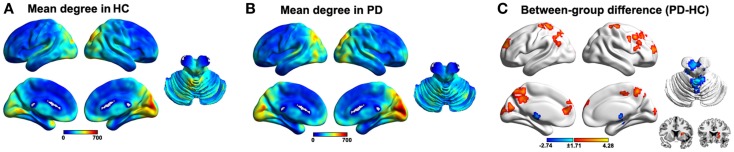
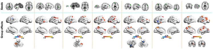
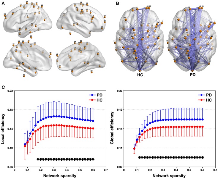
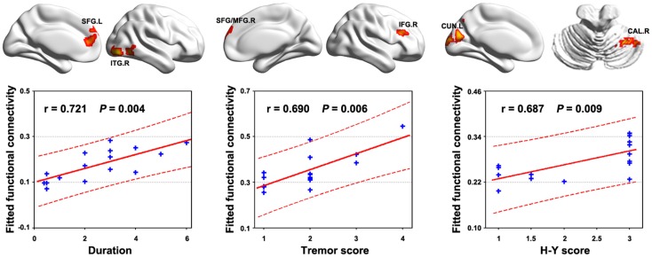
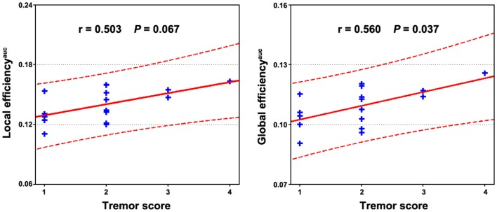
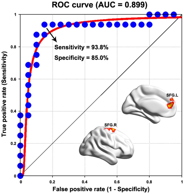
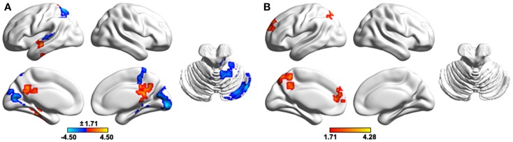
Parkinson's disease (PD) is a clinically heterogeneous disease in the symptomatology dominated by tremor, akinesia, or rigidity. Focusing on PD patients with tremor, this study investigated their discoordination patterns of spontaneous brain activity by combining voxel-wise centrality, seed-based functional connectivity, and network efficiency methods. Sixteen patients and 20 matched healthy controls (HCs) were recruited and underwent structural and resting-state functional MRI scan. Compared with the HCs, the patients exhibited increased centrality in the frontal, parietal, and occipital regions while decreased centrality in the cerebellum anterior lobe and thalamus. Seeded at these regions, a distributed network was further identified that encompassed cortical (default mode network, sensorimotor cortex, prefrontal and occipital areas) and subcortical (thalamus and basal ganglia) regions and the cerebellum and brainstem. Graph-based analyses of this network revealed increased information transformation efficiency in the patients. Moreover, the identified network correlated with clinical manifestations in the patients and could distinguish the patients from HCs. Morphometric analyses revealed decreased gray matter volume in multiple regions that largely accounted for the observed functional abnormalities. Together, these findings provide a comprehensive view of network disorganization in PD with tremor and have important implications for understanding neural substrates underlying this specific type of PD.
Keywords: Parkinson’s disease; centrality; connectome; resting functional connectivity; tremor.
Figures
Similar articles
-
Altered resting-state voxel-level whole-brain functional connectivity in depressed Parkinson's disease.Parkinsonism Relat Disord. 2018 May;50:74-80. doi: 10.1016/j.parkreldis.2018.02.019. Epub 2018 Feb 9. Parkinsonism Relat Disord. 2018. PMID: 29449183
-
Increased thalamic centrality and putamen-thalamic connectivity in patients with parkinsonian resting tremor.Brain Behav. 2016 Nov 23;7(1):e00601. doi: 10.1002/brb3.601. eCollection 2017 Jan. Brain Behav. 2016. PMID: 28127519 Free PMC article.
-
Disrupted Functional Connectivity of Basal Ganglia across Tremor-Dominant and Akinetic/Rigid-Dominant Parkinson's Disease.Front Aging Neurosci. 2017 Nov 2;9:360. doi: 10.3389/fnagi.2017.00360. eCollection 2017. Front Aging Neurosci. 2017. PMID: 29163141 Free PMC article.
-
Mapping Essential Tremor to a Common Brain Network Using Functional Connectivity Analysis.Neurology. 2023 Oct 10;101(15):e1483-e1494. doi: 10.1212/WNL.0000000000207701. Epub 2023 Aug 18. Neurology. 2023. PMID: 37596042 Free PMC article. Review.
-
Oscillation-specific nodal alterations in early to middle stages Parkinson's disease.Transl Neurodegener. 2019 Nov 15;8:36. doi: 10.1186/s40035-019-0177-5. eCollection 2019. Transl Neurodegener. 2019. PMID: 31807287 Free PMC article. Review.
Cited by
-
Acupuncture Modulates Disrupted Whole-Brain Network after Ischemic Stroke: Evidence Based on Graph Theory Analysis.Neural Plast. 2020 Aug 19;2020:8838498. doi: 10.1155/2020/8838498. eCollection 2020. Neural Plast. 2020. PMID: 32922447 Free PMC article.
-
Multidelay multiparametric arterial spin labeling perfusion MRI and mild cognitive impairment in early stage Parkinson's disease.Hum Brain Mapp. 2019 Mar;40(4):1317-1327. doi: 10.1002/hbm.24451. Epub 2018 Dec 7. Hum Brain Mapp. 2019. PMID: 30548099 Free PMC article.
-
Altered brain functional network in children with type 1 Gaucher disease: a longitudinal graph theory-based study.Neuroradiology. 2019 Jan;61(1):63-70. doi: 10.1007/s00234-018-2104-3. Epub 2018 Oct 8. Neuroradiology. 2019. PMID: 30298188
-
Graph theory analysis of resting-state functional magnetic resonance imaging in essential tremor.Hum Brain Mapp. 2019 Nov 1;40(16):4686-4702. doi: 10.1002/hbm.24730. Epub 2019 Jul 22. Hum Brain Mapp. 2019. PMID: 31332912 Free PMC article.
-
Predicting poststroke dyskinesia with resting-state functional connectivity in the motor network.Neurophotonics. 2023 Apr;10(2):025001. doi: 10.1117/1.NPh.10.2.025001. Epub 2023 Apr 4. Neurophotonics. 2023. PMID: 37025568 Free PMC article.
References
LinkOut - more resources
Full Text Sources
Other Literature Sources