Review
. 2015 Feb 3:6:12.
doi: 10.3389/fphys.2015.00012. eCollection 2015.

The regulation of muscle mass by endogenous glucocorticoids

Affiliations
Review

The regulation of muscle mass by endogenous glucocorticoids

Theodore P Braun et al. Front Physiol. .

Abstract

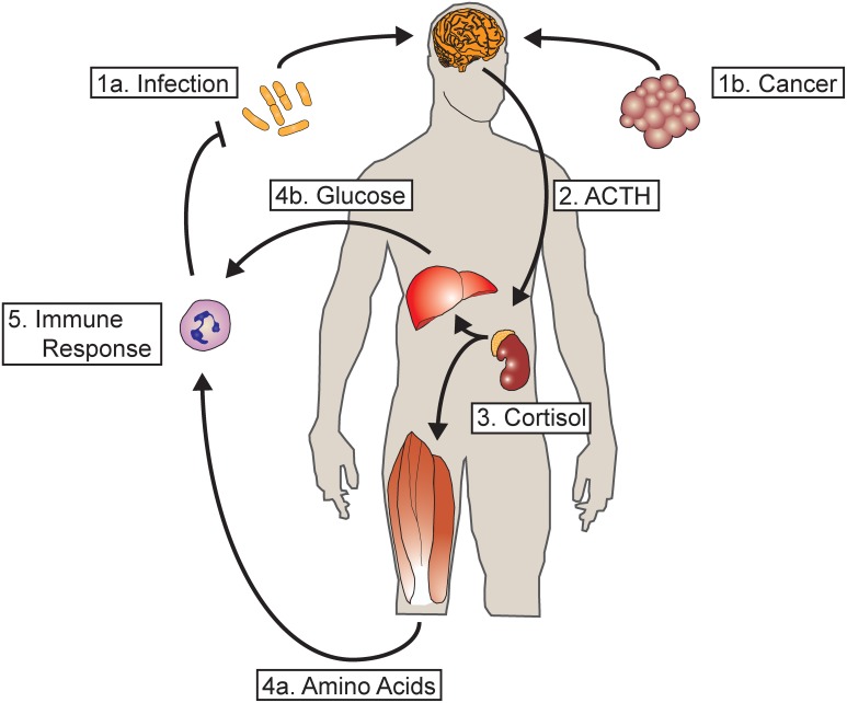
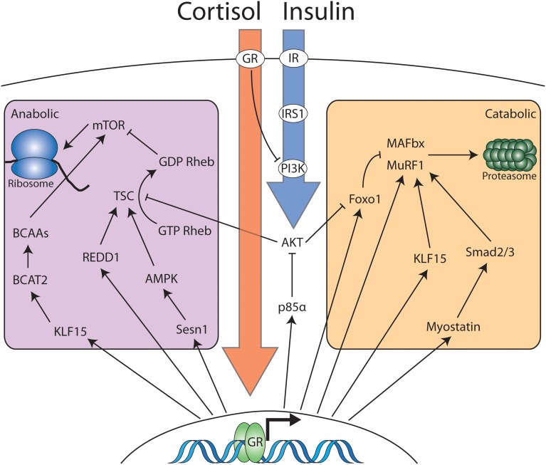
Glucocorticoids are highly conserved fundamental regulators of energy homeostasis. In response to stress in the form of perceived danger or acute inflammation, glucocorticoids are released from the adrenal gland, rapidly mobilizing energy from carbohydrate, fat and protein stores. In the case of inflammation, mobilized protein is critical for the rapid synthesis of acute phase reactants and an efficient immune response to infection. While adaptive in response to infection, chronic mobilization can lead to a profound depletion of energy stores. Skeletal muscle represents the major body store of protein, and can become substantially atrophied under conditions of chronic inflammation. Glucocorticoids elicit the atrophy of muscle by increasing the rate of protein degradation by the ubiquitin-proteasome system and autophagy lysosome system. Protein synthesis is also suppressed at the level of translational initiation, preventing the production of new myofibrillar protein. Glucocorticoids also antagonize the action of anabolic regulators such as insulin further exacerbating the loss of protein and muscle mass. The loss of muscle mass in the context of chronic disease is a key feature of cachexia and contributes substantially to morbidity and mortality. A growing body of evidence demonstrates that glucocorticoid signaling is a common mediator of wasting, irrespective of the underlying initiator or disease state. This review will highlight fundamental mechanisms of glucocorticoid signaling and detail the mechanisms of glucocorticoid-induced muscle atrophy. Additionally, the evidence for glucocorticoids as a driver of muscle wasting in numerous disease states will be discussed. Given the burden of wasting diseases and the nodal nature of glucocorticoid signaling, effective anti-glucocorticoid therapy would be a valuable clinical tool. Therefore, the progress and potential pitfalls in the development of glucocorticoid antagonists for muscle wasting will be discussed.

Keywords: HPA-axis; cachexia; catabolism; glucococorticoids; muscle; protein synthesis; skeletal; wasting.


Figures

Figure 1
Figure 1
Glucocorticoids mediate a metabolic response to acute and chronic inflammation. (1a) During acute infection, inflammatory mediators act in the central nervous system activating hypothalamic CRH neurons. (1b) Chronic inflammation, such as from tumor growth, also activates hypothalamic CRH neurons. (2) The release of CRH into the pituitary portal vasculature results in the release of ACTH by the anterior pituitary into the systemic circulation. (3) ACTH acts on the adrenal gland, resulting in the release of cortisol (or corticosterone in rodents). (4a) Cortisol acts on skeletal muscle, resulting in the breakdown of contractile protein and the mobilization of amino acids. (4b) Cortisol also acts on the liver to increase hepatic gluconeogenesis. (5) The resultant substrate mobilized from energy reserves serves to fuel the synthesis of acute phase reactants as well as the innate and adaptive immune response. In the case of acute infection, the resultant feedback loop results in resolution and clearance of the offending organism, terminating the inflammatory response and the mobilization of resources. However, in cases of chronic inflammation such as cancer where the immune response is unable to terminate the underlying inflammatory stimulus, continuous mobilization of amino acids leads to a profound depletion of skeletal muscle.
Figure 2
Figure 2
Cortisol and insulin signaling interact to modulate the anabolic and catabolic balance in skeletal muscle. Cortisol and corticosterone interact with cytosolic glucocorticoid receptor in skeletal muscle. The majority of the effects of cortisol occur via glucocorticoid receptor dimerization, nuclear localization and activation of transcription. However, cytosolic ligand bound GR monomers also antagonize insulin signaling at the level of PI3K. Nuclear GR induces the transcription of multiple genes that partition into two distinct domains: Genes that increase catabolic processes and genes that decrease anabolic processes. Increased anabolic activity occurs via increased ubiquitin-proteasome activity or increases in the activity of the autophagy-lysosome system (not pictured). The reduction in anabolic activity occurs via a number of pathways that converge to inhibit mTOR-dependent protein translation. Insulin signaling opposes glucocorticoid dependent mobilization of muscle protein via interaction with membrane bound insulin receptor. Signaling downstream of the insulin receptor results in inhibition of proteasomal targeting as well as stimulating mTOR-dependent protein translation.

References

    1. Akita S., Webster J., Ren S. G., Takino H., Said J., Zand O., et al. . (1995). Human and murine pituitary expression of leukemia inhibitory factor. Novel intrapituitary regulation of adrenocorticotropin hormone synthesis and secretion. J. Clin. Invest. 95, 1288–1298. 10.1172/JCI117779 - DOI - PMC - PubMed
    1. Allen D. L., Loh A. S. (2011). Posttranscriptional mechanisms involving microRNA-27a and b contribute to fast-specific and glucocorticoid-mediated myostatin expression in skeletal muscle. Am. J. Physiol. Cell Physiol. 300, C124–C137. 10.1152/ajpcell.00142.2010 - DOI - PMC - PubMed
    1. Almeida M., Han L., Ambrogini E., Weinstein R. S., Manolagas S. C. (2011). Glucocorticoids and tumor necrosis factor α increase oxidative stress and suppress Wnt protein signaling in osteoblasts. J. Biol. Chem. 286, 44326–44335. 10.1074/jbc.M111.283481 - DOI - PMC - PubMed
    1. Baehr L. M., Furlow J. D., Bodine S. C. (2011). Muscle sparing in muscle RING finger 1 null mice: response to synthetic glucocorticoids. J. Physiol. (Lond). 589, 4759–4776. 10.1113/jphysiol.2011.212845 - DOI - PMC - PubMed
    1. Baracos V., Rodemann H. P., Dinarello C. A., Goldberg A. L. (1983). Stimulation of muscle protein degradation and prostaglandin E2 release by leukocytic pyrogen (interleukin-1). A mechanism for the increased degradation of muscle proteins during fever. N. Engl. J. Med. 308, 553–558. 10.1056/NEJM198303103081002 - DOI - PubMed

LinkOut - more resources