

Comparison of human adipose-derived stem cells and chondroitinase ABC transplantation on locomotor recovery in the contusion model of spinal cord injury in rats
- PMID: 25691946
- PMCID: PMC4322153
Comparison of human adipose-derived stem cells and chondroitinase ABC transplantation on locomotor recovery in the contusion model of spinal cord injury in rats
Abstract
Objectives: Spinal cord injury (SCI) is one of the most serious clinical diseases and its treatment has been a subject of interest to researchers. There are two important therapeutic strategies in the treatment of SCI: replacing lost tissue cells through cells implantation and scar elimination. Therefore, in this study we used human adipose-derived stem cells (hADSCs) implantation and injection of Chondroitinase ABC. Aim of present study was to answer to this question: which one is more efficient for Improvement of locomotor recovery after SCI in rat? Transplantation of hADSCs or injection of ChABC.
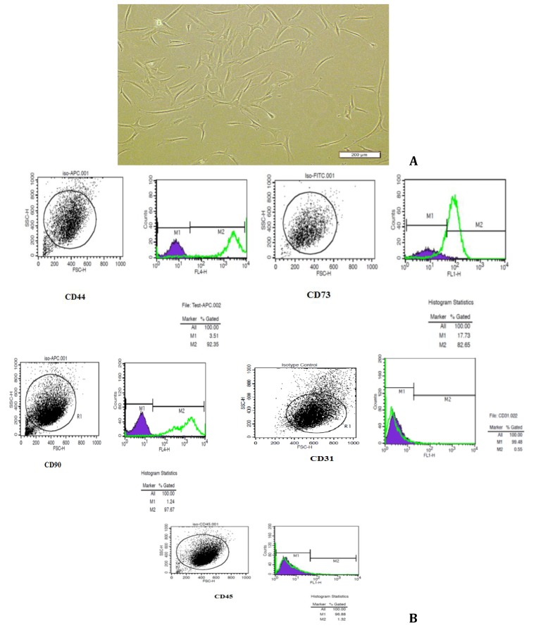
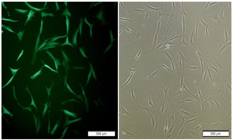
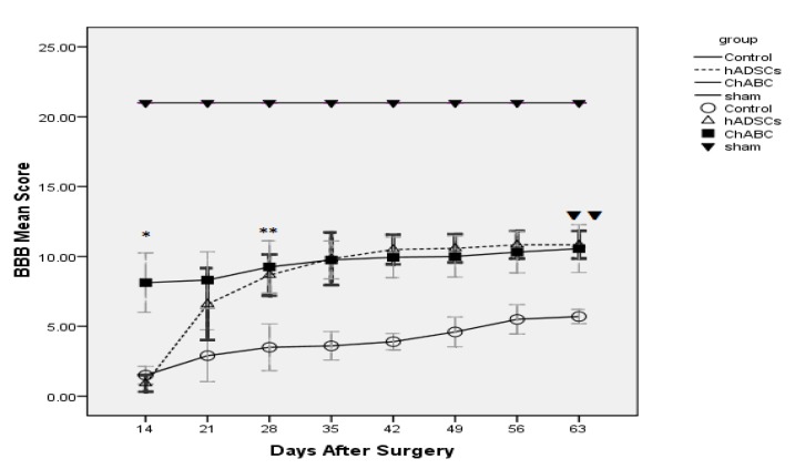
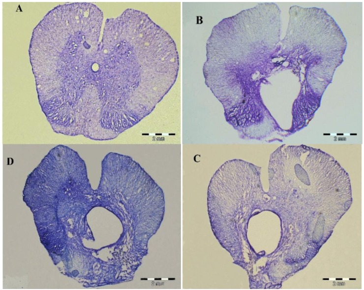
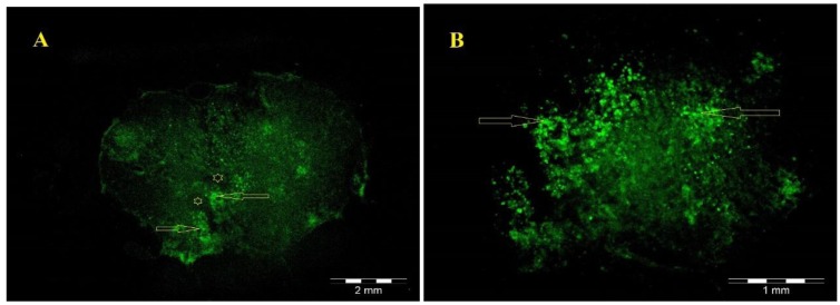
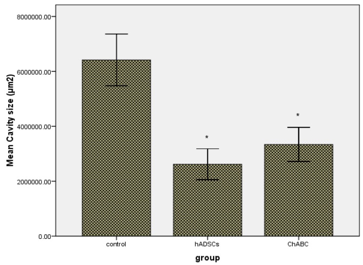
Materials and methods: The spinal cord of rats was injured by contusion using a weight-drop at the level of T8-9, the hADSCs and Chondroitinase ABC were infused in to the spinal cord tissue after injury. BBB test was performed and recorded for each animal weekly for 8 weeks. After the 8(th) weeks, Serial cross-sections were stained with cresyl violet and examined under a light microscope and area of cavity in the spinal cord was measured.
Results: At 8(th) weeks after injection, hADSCs and ChABC significantly promote locomotor function (P<0.01) and spinal cords of hADSCs and ChABC group had cavities much smaller than those of the control group (P<0.001).
Conclusion: Results of the present study shows dealing with inappropriate neuro-inhibitory environment and glial scar by ChABC have equal role compare to cell therapy (with hADSCs) for improving motor function after SCI and this result in adoption of proper therapeutic strategies for SCI intervention is important.
Keywords: BBB; Chondroitinase ABC; Contusion; Spinal Cord Injury; hADSCs.
Figures
Similar articles
-
The combined application of human adipose derived stem cells and Chondroitinase ABC in treatment of a spinal cord injury model.Neuropeptides. 2017 Feb;61:39-47. doi: 10.1016/j.npep.2016.07.004. Epub 2016 Jul 28. Neuropeptides. 2017. PMID: 27484347
-
[TRANSPLANTATION OF NEURAL STEM CELLS INDUCED BY ALL-TRANS- RETINOIC ACID COMBINED WITH GLIAL CELL LINE DERIVED NEUROTROPHIC FACTOR AND CHONDROITINASE ABC FOR REPAIRING SPINAL CORD INJURY OF RATS].Zhongguo Xiu Fu Chong Jian Wai Ke Za Zhi. 2015 Aug;29(8):1009-15. Zhongguo Xiu Fu Chong Jian Wai Ke Za Zhi. 2015. PMID: 26677625 Chinese.
-
Combined treatment with enteric neural stem cells and chondroitinase ABC reduces spinal cord lesion pathology.Stem Cell Res Ther. 2021 Jan 6;12(1):10. doi: 10.1186/s13287-020-02031-9. Stem Cell Res Ther. 2021. PMID: 33407795 Free PMC article.
-
Chondroitinase ABC Administration in Locomotion Recovery After Spinal Cord Injury: A Systematic Review and Meta-analysis.Basic Clin Neurosci. 2022 Sep-Oct;13(5):609-624. doi: 10.32598/bcn.2021.1422.1. Epub 2022 Sep 1. Basic Clin Neurosci. 2022. PMID: 37313020 Free PMC article. Review.
-
Combination treatment with chondroitinase ABC in spinal cord injury--breaking the barrier.Neurosci Bull. 2013 Aug;29(4):477-83. doi: 10.1007/s12264-013-1359-2. Epub 2013 Jul 9. Neurosci Bull. 2013. PMID: 23839053 Free PMC article. Review.
Cited by
-
[Effects of human urine-derived stem cells combined with chondroitinase ABC on the expressions of nerve growth factor and brain-derived neurotrophic factor in the spinal cord injury].Zhongguo Xiu Fu Chong Jian Wai Ke Za Zhi. 2017 Nov 15;31(11):1377-1383. doi: 10.7507/1002-1892.201706082. Zhongguo Xiu Fu Chong Jian Wai Ke Za Zhi. 2017. PMID: 29798595 Free PMC article. Chinese.
-
Transplantation of Neural Stem Cells Cultured in Alginate Scaffold for Spinal Cord Injury in Rats.Asian Spine J. 2016 Aug;10(4):611-8. doi: 10.4184/asj.2016.10.4.611. Epub 2016 Aug 16. Asian Spine J. 2016. PMID: 27559438 Free PMC article.
-
Stem cell transplantation and functional recovery after spinal cord injury: a systematic review and meta-analysis.Anat Cell Biol. 2018 Sep;51(3):180-188. doi: 10.5115/acb.2018.51.3.180. Epub 2018 Sep 28. Anat Cell Biol. 2018. PMID: 30310710 Free PMC article.
-
Laparoscopic adipose-derived stem cell harvest technique with bipolar sealing device: Outcome in 12 dogs.Vet Med Sci. 2022 Jul;8(4):1421-1428. doi: 10.1002/vms3.816. Epub 2022 May 10. Vet Med Sci. 2022. PMID: 35537084 Free PMC article.
-
Posterior Tibial Nerve Stimulation in Fecal Incontinence: A Systematic Review and Meta-Analysis.Basic Clin Neurosci. 2019 Sep-Oct;10(5):419-431. doi: 10.32598/bcn.9.10.290. Epub 2019 Sep 1. Basic Clin Neurosci. 2019. PMID: 32284831 Free PMC article.
References
-
- Pearse DD, Sanchez AR, Pereira FC, Andrade CM, Puzis R, Pressman Y, et al. Transplantation of Schwann cells and/or olfactory ensheathing glia into the contused spinal cord: Survival migration axon association nd functional recovery. Glia . 2007;55:976–1000. - PubMed
-
- Hulsebosch CE. Recent advances in pathophysiology and treatment of spinal cord injury. Adv Physiol Educ. 2002; 26:238–255. - PubMed
-
- Dietz V, Curt A. Neurological aspects of spinal-cord repair: promises and challenges. Lancet neurol . 2006;5:688–694. - PubMed
-
- McDonald JW, Belegu V. Demyelination and remyelination after spinal cord injury. J Neurotrauma. 2006; 23:345–359. - PubMed
LinkOut - more resources
Full Text Sources