

Lacunar infarction and small vessel disease: pathology and pathophysiology
- PMID: 25692102
- PMCID: PMC4325635
- DOI: 10.5853/jos.2015.17.1.2
Lacunar infarction and small vessel disease: pathology and pathophysiology
Abstract
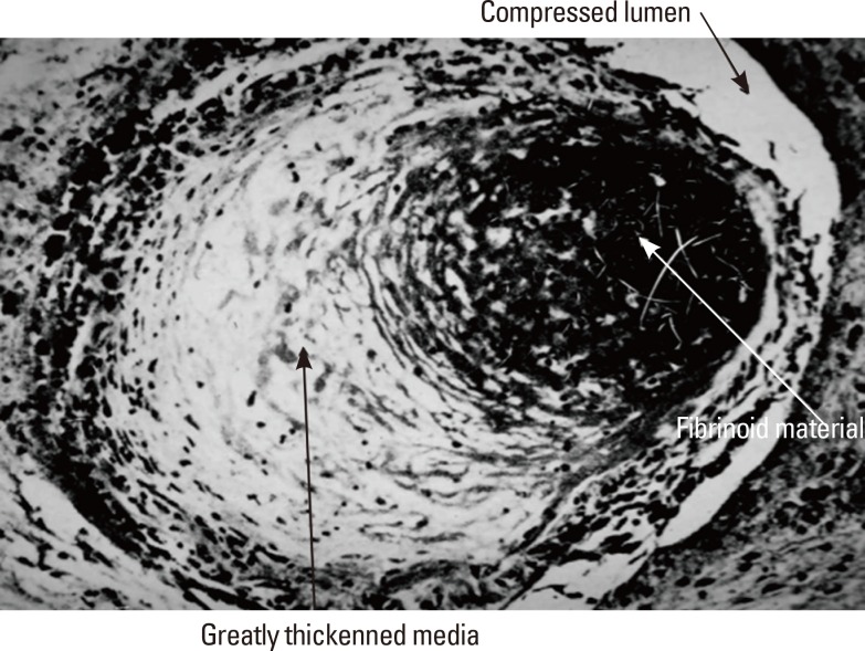
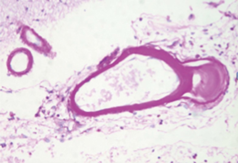
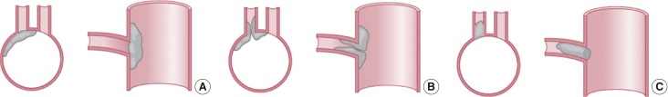
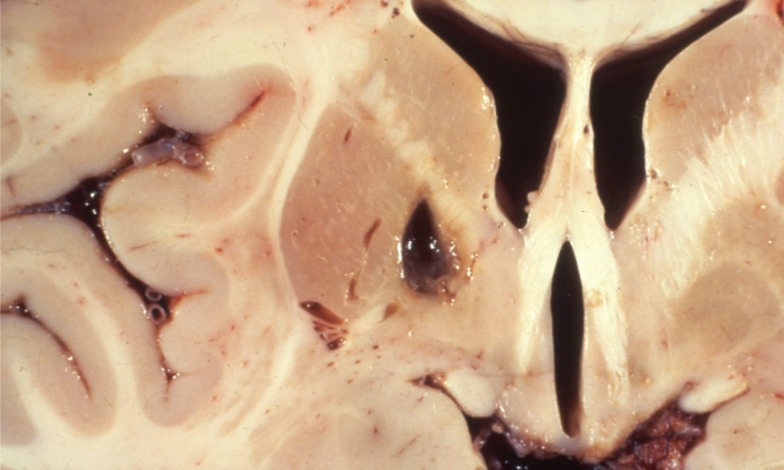
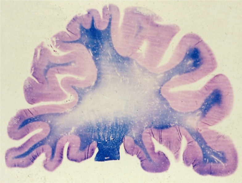
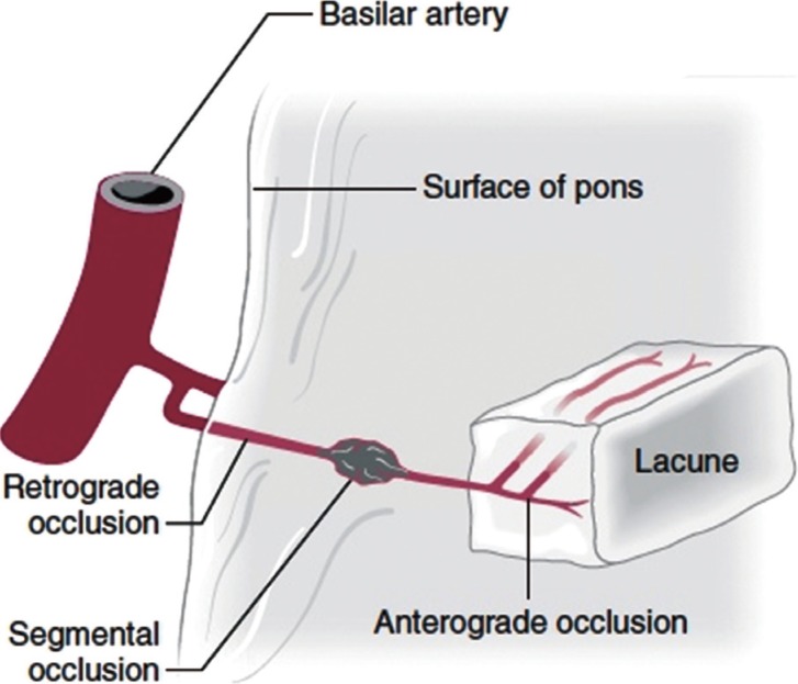
Two major vascular pathologies underlie brain damage in patients with disease of small size penetrating brain arteries and arterioles; 1) thickening of the arterial media and 2) obstruction of the origins of penetrating arteries by parent artery intimal plaques. The media of these small vessels may be thickened by fibrinoid deposition and hypertrophy of smooth muscle and other connective tissue elements that accompanies degenerative changes in patients with hypertension and or diabetes or can contain foreign deposits as in amyloid angiopathy and genetically mediated conditions such as cerebral autosomal dominant arteriopathy with subcortical infarcts and leukoencephalopathy. These pathological changes lead to 2 different pathophysiologies: 1) brain ischemia in regions supplied by the affected arteries. The resultant lesions are deep small infarcts, most often involving the basal ganglia, pons, thalami and cerebral white matter. And 2) leakage of fluid causing edema and later gliosis in white matter tracts. The changes in the media and adventitia effect metalloproteinases and other substances within the matrix of the vessels and lead to abnormal blood/brain barriers in these small vessels. and chronic gliosis and atrophy of cerebral white matter.
Keywords: CADASIL; Cerebral amyloid angiopathy; Cerebral small vessel diseases; Pathophysiology.
Conflict of interest statement
The authors have no financial conflicts of interest.
Figures
References
-
- Fisher CM. The arterial lesions underlying lacunes. Acta Neuropathol. 1968;12:1–15. - PubMed
-
- Fisher CM. Lacunes, small deep cerebral infarcts. Neurology. 1965;15:774–784. - PubMed
-
- Fisher CM. Binswanger's encephalopathy: A review. J Neurol. 1989;236:65–79. - PubMed
-
- Chabriat H, Joutel A, Dichgans M, Tournier-Lasserve E, Bousser MG. CADASIL. Lancet Neurol. 2009;8:643–653. - PubMed
Publication types
LinkOut - more resources
Full Text Sources
Other Literature Sources
Medical
